Your Visit | Overview
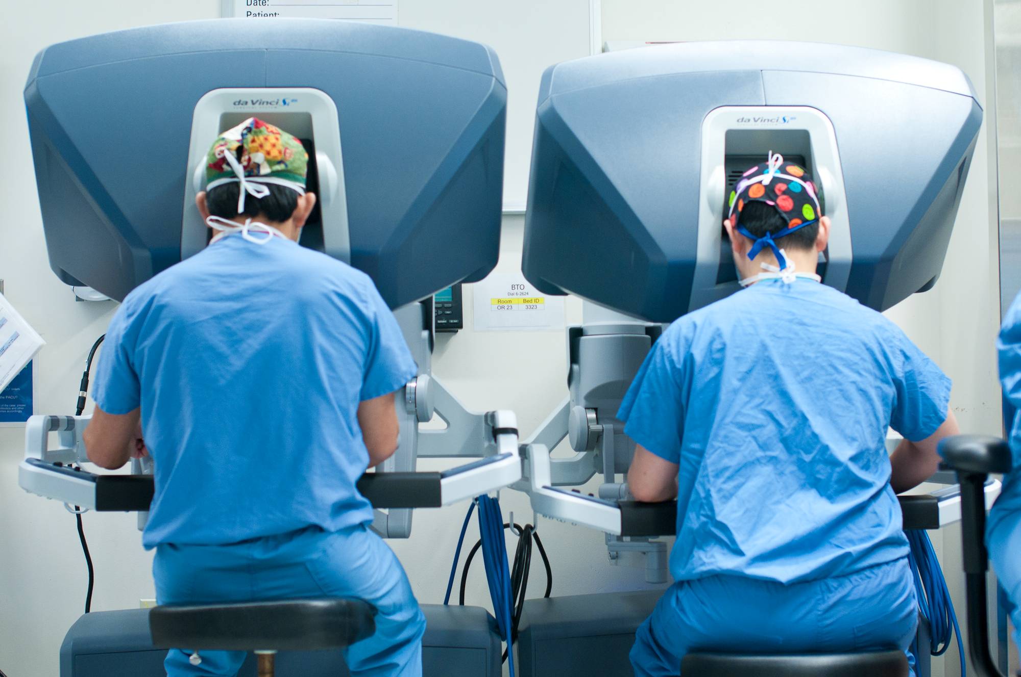 The Robotic Surgery Research and Training Program at Boston Children's Hospital believes every child is entitled to the most technologically advanced care possible delivered by a collaborative team of experts. We employ the latest advances in computerized diagnostics and treatment techniques. This, combined with the clinical skills of the medical staff, ensures the highest level of patient care.
Common procedures performed at Boston Children’s Hospital include:
Pyeloplasty
A pyeloplasty procedure repairs ureteropelvic junction obstruction (UPJ), the most common type of blockage that causes hydronephrosis (build-up of fluid in the kidney).
There are two methods of pyeloplasty used at Boston Children’s: laparoscopy or robot-assisted laparoscopy. Both are minimally invasive procedures, but the robot-assisted approach allows surgeons to have better control of the instruments so they can more easily perform complex maneuvers. About half of the pyeloplasties performed at Boston Children’s are robot-assisted laparoscopy procedures.
During a robot-assisted pyeloplasty, very thin instruments are inserted into three or four small incisions. Operating with the aid of a tiny camera, surgeons remove the narrowed or obstructed part of the ureter (the tube connecting the bladder to the kidney) and reconnect the healthy portion to the kidney’s drainage system.
The success rate of pyeloplasty procedures is about 95 percent. After a robot-assisted pyeloplasty, children are usually able to go home the following day.
Mitrofanoff procedure
Also known as appendo-vesicostomy, the Mitrofanoff procedure offers an alternative to catheterization through the urethra by using a surgically-created catheterizable channel. The channel goes from the bladder to the abdominal wall, usually through the belly button.
Bilateral ureteral reimplantation
Bilateral ureteral reimplantation is a surgical procedure used to treat vesicoureteral reflux (VUR), a condition in which the urine flows backwards from the bladder up toward the kidneys. VUR can put children at risk for kidney infections, which may lead to kidney damage.
During the procedure, the surgeon makes an incision in the lower abdomen and exposes the bladder. The junction of the bladder and the ureter (the tube connecting the bladder to the kidney) is reconstructed to prevent urine from flowing backward up into the kidney. A catheter is left in the bladder to drain the urine for the first one to two days after surgery.