What is double outlet right ventricle?
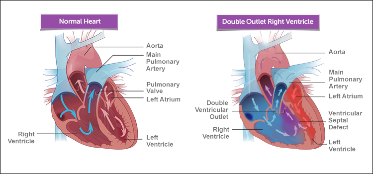
Double outlet right ventricle (DORV) is a rare congenital heart defect, meaning it’s a condition a baby is born with. In DORV, the pulmonary artery and the aorta — the heart’s two major arteries — both connect to the right ventricle. In a normal heart, the pulmonary artery connects to the right ventricle, and the aorta connects to the left ventricle. DORV creates a problem because the right ventricle carries oxygen-poor blood, which then gets circulated in the body.
Another heart condition, called a ventricular septal defect (VSD), always occurs with DORV. This is a hole in the tissue wall (septum) that normally separates the right and left ventricles. The VSD allows oxygen-rich blood to pass from the left ventricle to the aorta and pulmonary artery. But even with this added oxygen, the body may still not get enough, causing the heart to work harder.
A child with DORV may also have other heart problems, including:
- pulmonary (valve) stenosis (PVS, PS)
- transposition of the great arteries (TGA)
- pulmonary atresia
- coarctation of the aorta
- mitral valve abnormalities
If your child has DORV, the severity of the condition and type of treatment, including the type of surgical repair, will vary depending on which types of defects he or she has. Although DORV is a serious condition, it is treatable with surgery.
What are the symptoms of double outlet right ventricle?
Common symptoms of double outlet right ventricle (DORV) in babies include:
- rapid breathing
- rapid heartbeat
- sweating
- disinterest in feeding or tiring while feeding
- poor weight gain
- blue color of the skin, lips and nail beds (cyanosis)
- heart murmur (detected by doctor)
In older children, symptoms may include:
- fatigue
- shortness of breath
How we care for double outlet right ventricle
The experienced surgeons in the Boston Children’s Hospital Cardiac Surgery Department treat some of the most complex pediatric heart conditions in the world, with overall success rates approaching 98 percent — among the highest in the nation among large pediatric cardiac centers. The success rates of the operations used to repair most forms of DORV are in the same range.
At Boston Children’s, we provide families with a wealth of information, resources, programs and support — before, during and after your child’s treatment. With our compassionate, family-centered approach to expert treatment and care, you and your child are in the best possible hands.
DORV | Diagnosis & Treatments
How is double outlet right ventricle diagnosed?
If your newborn baby has a bluish tint to the skin or your young child has symptoms of a heart condition, your pediatrician will refer you to a pediatric cardiologist (heart doctor) for a full examination.
The cardiologist will listen to your baby’s heart and lungs to assess for a heart murmur — a noise heard through a stethoscope that is caused by the turbulence of blood flow. He or she will also measure the oxygen level in your baby’s blood. This examination will give the doctor an idea of the kind of heart problem your baby may have.
In some cases, signs of a congenital heart defect may be found by a routine prenatal ultrasound during pregnancy. If this is the case, a cardiac ultrasound of the baby in utero is usually the next step.
The doctor may also order one or more of the following tests to diagnose DORV and any related heart conditions:
- electrocardiogram(ECG or EKG)
- echocardiogram (cardiac ultrasound)
- chest x-ray
- cardiac catheterization
What are the treatment options for double outlet right ventricle?
Children with double outlet right ventricle (DORV) will need surgery to close the hole in the heart and connect the aorta to the left ventricle and the pulmonary artery to the right ventricle.
The most common surgical procedure for DORV with a ventricular septal defect (VSD) is called intraventricular tunnel repair. For this surgery, the surgeon creates a kind of tunnel to connect the left ventricle to the aorta. A patch placed within the heart directs left ventricle blood flow to the aorta.
Another procedure, called an arterial switch operation, is necessary if the aorta and the pulmonary artery are reversed in relation to each other. For this procedure, a tunnel from the VSD to the pulmonary artery is created, connecting the left ventricle to the pulmonary artery. Then, the vessels are disconnected and reconnected so that the pulmonary artery becomes the aorta and the aortic valve is connected to the pulmonary artery, and the holes between the left and right ventricles of the heart are closed.
If your child has other heart conditions associated with DORV, he or she may also need other types of surgery.
Make an appointment
- Call 617-355-4278
- Heart@childrens.harvard.edu
- Learn more about the Complex Biventricular Repair Program
- If you are pregnant, please visit the Fetal Cardiology Program
- Request a second opinion