Age-Related Macular Degeneration | Overview
Anatomical Background of AMD
- Age-related macular degeneration (AMD) is the leading cause of vision loss in adults over 50. It can occur in two forms, wet AMD and dry AMD.
- In dry AMD, cellular debris called drusen accumulates between the retina and the choroid, causing damage to the retina.
- In wet AMD, blood vessels infiltrate from the retina to the choroid and cause hemorrhaging.
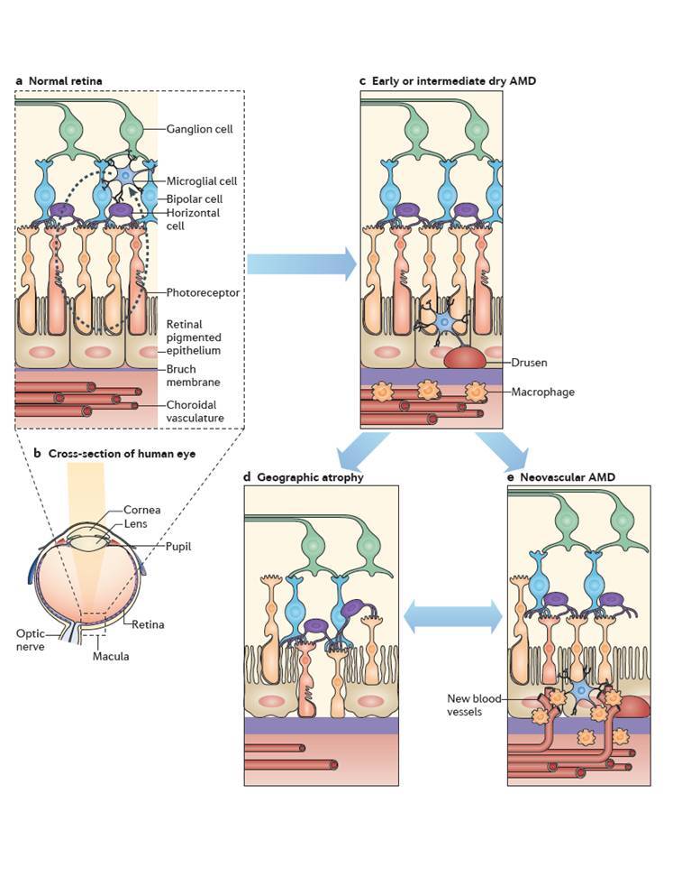
Above: A diagram illustrating the anatomical differences between a normal human retina (A,B) and a retina experiencing AMD. Early AMD involves the accumulation of drusen and macrophages in the subretinal space (E). This can then progress to either dry AMD (D) which is characterized by photoreceptor degeneration and regression of the choroid vasculature, or wet AMD (E) in which neovascularization from invading choroid vessels and macrophages rupture the Bruch’s membrane and cause photoreceptor damage. Source: Ambati, J., Atkinson, J. P., & Gelfand, B. D. (2013). Immunology of age-related macular degeneration. Nature Reviews Immunology, 13(6), 438-451.
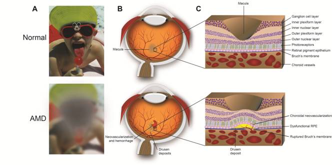
Below: How wet AMD affects vision. Neovascularization of the retina ruptures the Bruch’s membrane (C) which damages the macula (B) and results in blurry or spotty vision (A).
Choroidal Neovascularization
- Our lab uses a model of laser-induced choroidal neovascularization (CNV) to replicate wet AMD by rupturing the Bruch’s membrane and causing new blood vessels to develop in the choroid.
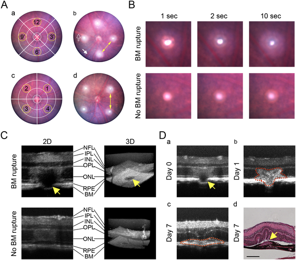
Above: Demonstration of suggested laser positions (A) and successful Bruch's Membrane (BM) rupture (B-D) in the mouse eye. Source: Gong Y, Li J, Sun Y, Fu Z, Liu C-H, Evans L, et al. (2015) Optimization of an Image-Guided Laser-Induced Choroidal Neovascularization Model in Mice. PLoS ONE 10(7): e0132643. doi:10.1371/journal.pone.0132643
- This is an excellent model to investigate the effect of different genes and treatments on vascular development because it can be used on any type of transgenic or knockout mice.
- Measuring the size of laser-induced lesions between different experimental groups allows for investigation of pro- and anti-inflammatory effects of treatments and genes.
- We recently published a paper demonstrating the proper procedure to use the Micron IV laser for the CNV model (Link).
- However this model is an injury model, rather than a true degenerative model which limits its effectiveness for investigating AMD.