What is a ventilation/perfusion scan?
A ventilation/perfusion scan is a diagnostic nuclear medicine test that is used to evaluate the circulation of air and blood within your child's lungs. The ventilation part of the test looks at the ability of air to reach all parts of the lungs, while the perfusion part evaluates how well the blood circulates within the lungs.
During the ventilation part of the test, a mask attached to a bag filled with oxygen and a radiopharmaceutical gas, called Xenon-133, will be placed over your child's nose and mouth. Your child will be asked to breathe normally into the bag.
During the perfusion part of the test, a radiopharmaceutical called Technetium-99m MAA will be injected in one of your child's veins. A special camera, called a gamma camera, is used to take pictures of the lungs after the radiopharmaceutical has been injected.
When might a ventilation/perfusion scan be needed?
 A ventilation/perfusion scan can help:
- evaluate disorders of your child's vascular system in the lungs (pulmonary embolism, infarct, thrombosis)
- make sure your child is all right after cardiac catheterization or cardiothoracic surgery
- evaluate children following congenital diaphragmatic hernia repair
- detect any blockage in your child's airway
How should I prepare my child for ventilation/perfusion scan?
There is no special preparation needed for this test.
- If a recent chest x-ray has not been performed, we may need to send you for one prior to the ventilation/perfusion scan.
- It is helpful to give your child a simple explanation as to why the scan is needed and assure her that you will be there for the entire time.
- We have various DVDs to choose from for your child to watch during the scan or you can bring one from home, although the test is only 10 minutes in length.
What should I expect when I bring my child to the hospital for a ventilation/perfusion scan?
When you arrive, please go to the Nuclear Medicine check-in desk on the second floor of the main hospital. A clinical intake coordinator will check in your child and verify her registration information.
What happens during a ventilation/perfusion scan?
You will be greeted by one of our nuclear medicine technologists who will explain to you and your child what will happen during the examination. The examination is done in two parts:
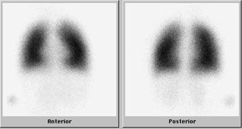
- ventilation: shows the flow of air in and out of the lungs
- perfusion: shows the blood flow to the lungs
Ventilation
- Imaging will take place during the entire ventilation phase of the exam.
- A mask attached to a bag filled with oxygen and a radiopharmaceutical gas will be placed over your child's nose and mouth. The gas is tasteless and odorless.
- Your child will be asked to breathe normally into the bag.
- At the end of that minute, a special exhaust fan will be placed near your child's head to help eliminate the exhaled gas.
Perfusion
- A radiopharmaceutical will be injected in one of your child's veins.
- Imaging of the lungs will be performed.
- It is important that your child remains as still as possible during imaging to obtain the best quality images.
Will my child feel anything during a ventilation/perfusion scan?
The placement of the mask over the mouth and nose may cause some anxiety or discomfort, but will not harm your child.
Your child may experience some discomfort associated with the insertion of the intravenous needle. The needle used for the procedure is small. Once the radiopharmaceutical is injected, the needle is withdrawn and a bandage is placed over the site of the injection. The area where the injection was given may be a little sore.
Although the gamma camera may appear large and intimidating, it does not touch your child.
Is a ventilation/perfusion scan safe?
We are committed to ensuring that your child receives the smallest radiation dose needed to obtain the desired result.
- Nuclear medicine has been used on babies and children for more than 40 years with no known adverse effects from the low doses employed.
- The radiopharmaceutical contains a very tiny amount of radioactive molecules. We believe that the benefit to your child's health outweighs potential radiation risk.
- The camera used to obtain the images does not produce any radiation.
- It is safe to be with your child during the procedure if you are pregnant or nursing.
What happens after the ventilation/perfusion scan?
Once the scan is complete, the images will be evaluated for quality. If the scan is adequate, your child will be free to leave and resume normal activity.
One of the Boston Children's nuclear medicine physicians will review your child's images and create a report of the findings and diagnosis.
How do I learn the results of the ventilation/perfusion scan?
The nuclear medicine physician will provide a report to the doctor who ordered your child's ventilation/perfusion scan. Your child's doctor will then discuss the results with you.
How Boston Children's Hospital approaches ventilation/perfusion scans
Our Nuclear Medicine and Molecular Imaging staff are committed to providing a safe, comfortable and child-friendly atmosphere with:
- specialized nuclear medicine physicians with expertise in interpreting ventilation/perfusion scans in children of all ages
- certified nuclear medicine technologists with years of experience imaging children and teens
- equipment adapted for pediatric use, which means age-appropriate care for children
- protocols that keep radiation exposure as low as reasonably achievable while assuring high image quality