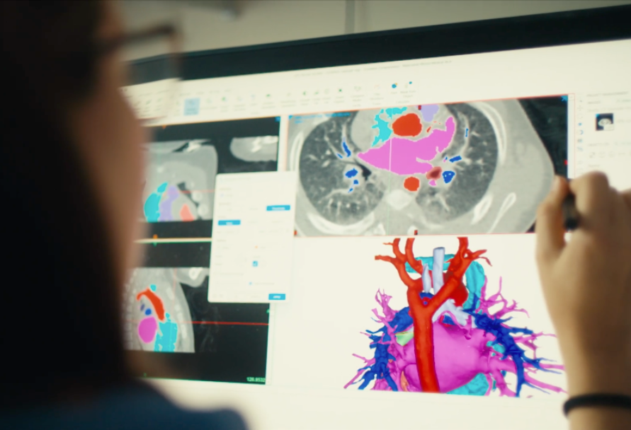
Surgical repair of a congenital heart defect (CHD) is a complex three-dimensional challenge. Every child is unique, and every CHD is different. Understanding the intricate details of each heart’s 3D anatomy to plan and conduct reconstructive surgeries that treat CHD is essential for an excellent outcome.
Historically, cardiac surgeons have relied on two-dimensional diagnostic information from echocardiograms, cardiac CT scans, and MRIs but had limited three-dimensional visualization of the heart. This left much of the burden on a surgeon to understand the 3D details of a patient’s heart anatomy only when they entered the operating room and then, in the moment, plan and conduct a perfect reconstruction.
Boston Children’s Cardiovascular 3D Modeling and Simulation Program has created and institutionalized a standard of preoperative planning that means our surgeons now rarely have to “figure it out” in the operating room. We have developed an exceptional team of engineers who are using the best available engineering software tools to create patient-specific 3D models of a child’s heart and blood vessels for preoperative planning and intraoperative guidance.
Check out our Paid Post video with The New York Times showing how our cardiac surgeons and engineers collaborate on 3D models that are helping drive the future of heart surgery — before a patient even enters the operating room.
Improving surgical planning and helping families understand the heart
Started in 2018, our program has since exploded in its breadth and impact. We have created more than 1,000 patient-specific 3D models. It is now a standard of care at the Benderson Family Heart Center for us to create 3D models and perform detailed analyses of a patient’s heart anatomy to help cardiac surgeons and cardiologists create intricate preoperative plans for many of the different CHD diagnoses that Boston Children’s treats.
The 3D models are shared at Heart Center surgical planning conferences and used as a tool to communicate among surgeons, cardiologists, anesthesiologists, fellows, and nurses. Surgical teams also consult models during an operation. The models also help families. As parents of a child who has complex CHD know, it’s challenging to understand the details of a heart anatomy from websites, cartoon drawings, or rudimentary sketches. We use the 3D models to provide incredible clarity, empowering families to understand cardiac disease and the Heart Center’s plans to repair it.
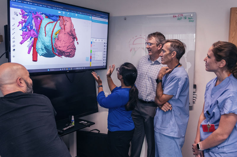
Learn how 3D modeling has transformed the planning of surgery by showing details of a patient’s heart.
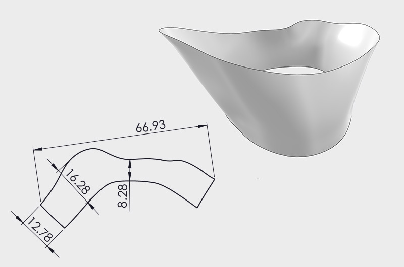
Understanding dimensions to plan ideal patches for repair
Built using a patient’s own imaging, these high-fidelity 3D models have accurate dimensions and spatial relationships. We use them to digitally plan complex repairs, including aortic and mitral valve reconstructions, ventricular septal defect (VSD) closures, and the augmentation of blood vessels (including pulmonary arteries and aortas).
Our engineering team works closely with advanced simulation companies across the world, utilizing engineering platforms developed for the aerospace and automotive industries to efficiently plan heart repair patches and complex reconstructions in our Heart Center. Visualizing the shape and size of an optimal patch is one thing, but accurately translating that information onto sterile patch material in the operating room (OR) is another. Inspired by the aerospace industry, we now use a laser system to project the shape of a designed patch onto patch material in the OR, where the design can be traced out and serve as a highly precise and sterile guide for a surgeon to complete the repair with unparalleled accuracy.
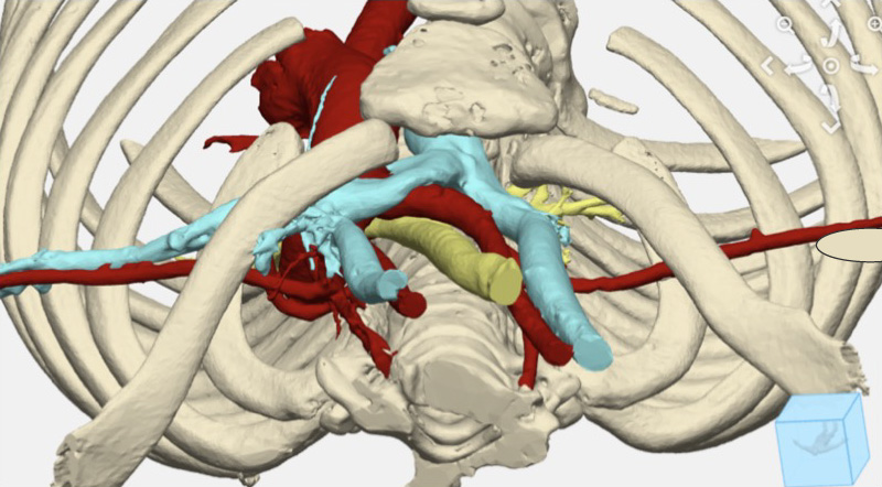
The intricate puzzle of chest wall reconstruction surgery was simplified with 3D modeling. See how it improved the treatment of complex cases.
Simulating blood flow for complex cases
In many cases, the complexity of heart disease includes so many variables that knowing exactly how to reconstruct the heart by intuition alone is impossible, even for the most experienced cardiac surgeons. The best distribution of blood flow with the best flow efficiency cannot be predicted just by clinical experience.
Our engineering team uses the same flow simulation software used to design aircraft to predict blood flow patterns within the heart and vessels after reconstruction. This tool has become indispensable in making decisions about some of our most complex cases, including children who have single ventricle defects and are undergoing Glenn and Fontan palliation.
This flow simulation, called computational fluid dynamics, can show surgeons and cardiologists the benefit of different surgical approaches, including where to place a Fontan conduit, how big to make it, and how to position it relative to Glenn connections to ensure balanced flow distribution with low energy loss to both lungs. Flow simulation has become our mainstay approach for managing complex single ventricle cases. We are the only Heart Center in the U.S. that performs this analysis as part of routine clinical care, and we also do it for patients at other heart centers around the world.
Technology used in aerospace is used to improve complex heart treatment. Read how aerospace engineering tools found a home at the Benderson Family Heart Center.
Setting a new standard in heart care
Our Cardiovascular 3D Modeling and Simulation Program is a unique aspect of the Benderson Family Heart Center. Our dynamic team of experienced engineers is leading the field in the development and implementation of advanced 3D modeling and simulation approaches to truly move the needle on how we care for patients with complex congenital heart disease.