3D Rendering of a Developing Mouse Embryo

Modeling the oculomotor system in mice: Whole mount imaging of a genetically modified mouse embryo with the motor neurons labeled in green and the muscles labeled in red allows us to trace the ocular motor nerves on their path from the brainstem to the orbit. Embryos are cleared using an iDisco protocol, imaged on a confocal microscope, and movies are produced using Imaris software.
New Publication!
New research shows that younger age and poor control at presentation but not magnitude of the deviation or stereopsis are associated with future surgery in intermittent exotropia.
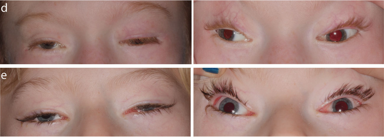
Whitman MC, Barry BJ, Robson CD, Facio FM, Van Ryzin C, Chan WM, Lehky TJ, Thurm A, Zalewski C, King KA, Brewer C, Almpani K, Lee JS, Delaney A, FitzGibbon EJ, Lee PR, Toro C, Paul SM, Abdul Rahman OA, Webb BD, Wang Jabs E, Moller HU, Ancher Larsen D, Antony JH, Troedson C, Ma A, Ragnhild G, Wirgenes KV, Tham E, Kvarnung M, Maarup TJ, MacKinnon S, Hunter DG, Collins FS, Manoli I, Engle EC. TUBB3 Arg262His causes a recognizable syndrome including CFEOM3, facial palsy, joint contractures, and early onset peripheral neuropathy. Human Genetics 2021 Online before print. Abstract
Microtubules form a cellular scaffold that is essential for proper development and function of the nervous system. Missense mutations in different tubulin isoforms cause brain malformations, and in TUBB3 can cause congenital fibrosis of the extraocular muscles type 3 (CFEOM3) and/or malformations of cortical development. We tested 14 individuals from 13 unrelated families who had a phenotype referred to as the TUBB3 R262H syndrome (the most severe form recognized to date). Affected individuals present at birth with ptosis, ophthalmoplegia, exotropia, facial weakness, facial dysmorphisms, and, in most cases, distal congenital joint contractures and other progressive disabilities before age 10. These subjects require multidisciplinary care, including ophthalmology, orthopedics/physiatry, physical therapy, neurology, and endocrinology. Care must be taken to prevent and treat corneal exposure and amblyopia to maximize vision, for example. These newly described phenotypes lead to better characterization of the spectrum and severity of disease associated with mutations in TUBB3, enhancing the accuracy of genetic testing and allowing for more personalized medical care and genetic counseling.
An article by Dr. Mary Whitman was featured in the Neuroscience section of the Journal of Visualized Experiments (JoVE). The video illustrates her original culturing technique, allowing identification of axon guidance pathways active in the oculomotor nerve and assessment of their roles at different points along the nerve trajectory in real time. Living slices are generated by embedding E10.5 IslMN:GFP embryos in agarose, slicing on a vibratome, and growing in a stage-top incubator.
Whitman MC, Bell JL, Nguyen EH, Engle EC. Ex Vivo Oculomotor Slice Culture from Embryonic GFP-Expressing Mice for Time-Lapse Imaging of Oculomotor Nerve Outgrowth. J Vis Exp (149), e59911, doi:10.3791/59911 (2019).
Dr. Whitman collaborated with a group that perfected the protocol to efficiently purify and cultivate ocular motor neurons and trochlear neurons alongside spinal motor neuron counterparts from the same embryos in transgenic mouse embryos.
Isolation and Culture of Oculomotor, Trochlear, and Spinal Motor Neurons from Prenatal Islmn: GFP Transgenic Mice. Fujiki R, Lee JY, Jurgens JA, Whitman MC, Engle EC. J Vis Exp 2019 Nov 12;(153):10.3791/60440. PMID: 31789317
In her ongoing study of oculomotor development, Dr. Mary Whitman (with Dr. Elizabeth Engle) published an article in the high-impact journal, Human Molecular Genetics. They report a new gene, ACKR3 (also known as CXCR7) that causes miswiring of the ocular motor nerves in humans and mice. Such miswiring leads to drooping eyelids and abnormal eye movements.
Whitman MC, Miyake N, Nguyen EH, Bell JL, Matos Ruiz PM, Chan WM, Di Gioia SA, Mukherjee N, Barry BJ, Bosley TM, Khan AO, Engle EC. Decreased ACKR3 (CXCR7) function causes oculomotor synkinesis in mice and humans. Hum Mol Genet. 2019 09 15; 28(18):3113-3125. PMID: 31211835.