 Every academic year, the Children’s Hospital Ophthalmology Foundation sponsors a Visiting Professor Series that brings in the best of the best in terms of basic research in ophthalmology. Funding includes a faculty dinner, all travel expenses, and an honorarium. Visiting professors give a lecture or two and, where appropriate, participate in a patient conference, then meet with faculty, followed by lunch with trainees. The activity not only gives our faculty and trainees access to these experts, it also allows the leaders in our field to experience firsthand the quality of training and research in the Boston Children’s Department of Ophthalmology.
The series has been held exclusively on Zoom since 2020, but one expects that to change in future. Please contact Eric Aho at Eric.Aho@childrens.harvard.edu if you would like more information about an upcoming lecture.
Academic year 2022-23
- Andreas Stahl (Sept. 21, 2022)
- Padmaja Sankaridurg (date not decided)
Academic year 2021-22
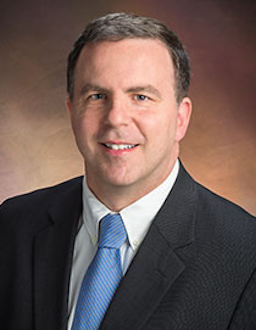 Dr. Avery is a pediatric neuro-ophthalmologist who spends 85% of his time performing clinical research and 15% seeing patients while actively teaching students, residents, and fellows. In his pediatric neuro-ophthalmology clinical practice, he is a member of numerous specialty centers within Children’s Hospital of Philadelphia (CHOP). He also established the Ophthalmology Ultrasound Specialty Clinic. He served as the lead ophthalmologist for a multi-disciplinary international task force to redefine the clinical criteria for NF1 and NF2, the former of which was recently published (Plotkin et al., Genetics in Medicine 2021). He serves as an Associate Editor for Neuropediatrics (journal for the German, Swiss, and Austrian Child Neurology Society) and Section Editor for the Journal of Neuro-Ophthalmology. He serves on the Quality Assurance and Low-Grade Glioma/Optic Pathway Glioma Committees and also chairs the Publication Committee for the Department of Defense Neurofibromatosis clinical trial consortium. He chairs the NIH/NCI (Response Evaluation in NF and Schwannomatosis) committee on OPGs. The primary goal of his clinical research program is to improve visual outcomes in children with OPGs. He spent the first five years of his career at Children’s National Medical Center in Washington, where he obtained his NEI K23 Career Development Award. He returned to CHOP/Penn during the last two years of his K23 while also becoming the PI of three foundation grants. Since then, he has developed a robust clinical research program where he is currently the PI on four federal grants spanning the National Eye Institute (R01), National Cancer Institute (UG3 and BIQSFP), and U.S. Department of Defense (Investigator Initiated Research Award), as well as being the PI for three active foundation grants. His OPG research has allowed him to participate in high-risk high-reward projects as part of the Gilbert Visual Restoration Initiative — a foundation funding cutting-edge research to produce visual restoration therapies in children with vision loss from their OPG secondary to NF1. Outside of his primary research focus, he remains active in publishing on ophthalmic imaging techniques to diagnose and monitor elevated ICP in children. Dr. Avery’s lectures were “Optic Pathway Gliomas: Clinical Challenges” and “Spasmus Nutans — A Neuro-Ophthalmic Syndrome of Poverty.”
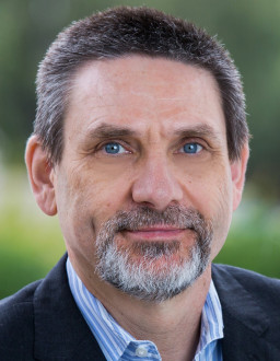 Dr. Palanker is a Professor of Ophthalmology and Electrical Engineering at Stanford University. He received a MSc in physics in 1984 from the State University of Armenia in Yerevan, and a PhD in applied physics in 1994 from the Hebrew University of Jerusalem.
Dr. Palanker studies interactions of electric field with biological cells and tissues, and develops optical and electronic technologies for diagnostic, therapeutic, surgical, and prosthetic applications, primarily in ophthalmology. In the range of optical frequencies, his studies include laser-tissue interactions with applications to ocular therapy and surgery, and interferometric imaging of the neural signals. In the field of electro-neural interfaces, he is working on high-resolution photovoltaic retinal prosthesis for restoration of sight and other implants for electronic control of organs. Several of his developments are in clinical practice worldwide: Pulsed Electron Avalanche Knife (PEAK PlasmaBlade), Patterned Scanning Laser Photocoagulator (PASCAL), Femtosecond Laser-assisted Cataract Surgery (Catalys), and Neural Stimulator for enhancement of the tear secretion (TrueTear). Photovoltaic retinal prosthesis for restoration of sight (PRIMA) is in clinical trials. His lecture was “Photovoltaic restoration of sight in age-related macular degeneration.”
Academic year 2020-21
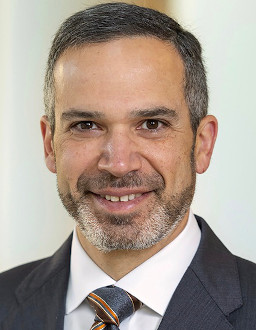 Dr. Kahana is a board-certified ophthalmologist and oculoplastic surgeon specializing in orbital disease and congenital eyelid conditions. A noted surgeon, scholar, author, and lecturer, Dr. Kahana spent the first 13 years of his career on faculty at the University of Michigan Kellogg Eye Center, where he rose to the rank of Associate Professor with Tenure, at which point he left to found the Michigan Center for Thyroid Eye Disease and Orbital Surgery. Dr. Kahana is a Professor of Ophthalmology at the Oakland University William Beaumont School of Medicine, an attending surgeon at the Children’s Hospital of Michigan, and a member of Consultants in Ophthalmic and Facial Plastic Surgery P.C. Dr. Kahana received his MD (with honors) as well as a PhD in molecular genetics and cell biology from The University of Chicago Pritzker School of Medicine. He completed his residency in ophthalmology and an ASOPRS fellowship in oculofacial plastic and reconstructive surgery at the University of Wisconsin. Dr. Kahana has earned an international reputation in orbital surgery — particularly thyroid eye disease and pediatric oculoplastics — particularly blepharophimosis and congenital ptosis. He has authored more than 70 peer-reviewed publications, multiple book chapters and reviews, and has given more than 100 lectures throughout the United States and internationally. Most recently he was honored to give the annual ASOPRS Foundation Lecture at the 2020 ASOPRS Spring Meeting. He has served in numerous leadership positions, including as President of the North American Society of Academic Orbital Surgeons (NASAOS) and Chair of the Thesis Committee and the Scientific Advisory Committee for ASOPRS. He is also a Program Director for his own ASOPRS fellowship. Dr. Kahana’s research has been awarded grants from the National Eye Institute of the National Institutes of Health, Research to Prevent Blindness, and other philanthropic and industry funding sources. Most recently, Dr. Kahana has been Principal Investigator of a major prospective clinical trial on the treatment of advanced orbital and periocular basal cell carcinoma. His presentations were “Bench to Bedside: The Biological Foundations of Pediatric Oculoplastic Surgery” and “Taking care of the parents and family of ophthalmic patients — Interesting Cases.”
 Professor Hellström is an outstanding researcher in the field of pediatric ophthalmology. She is world famous for her work on retinopathy of prematurity (ROP), a leading cause of childhood blindness. She has approached the research field of ROP using several techniques, developing innovative and reliable tools to optimize prediction of infants at risk, demonstrating important mechanisms of pathogenesis, discovering new, innovative therapies to prevent ROP, and evaluating these in clinical trials. Professor Hellström has published more than 200 papers in international, scientific journals and has an H-index of 53 with more than 11,000 citations. In a landmark paper, together with Professor Lois Smith published in PNAS in 2001, they showed in a mouse model that low IGF-1 suppresses VEGF-survival signaling in retinal endothelial cells and showed that this was directly correlated with the development of clinical ROP. The early results paved the way for a series of important papers, confirming that IGF-1 is critical for normal vascularization of the retina and that IGF-1 deficiency is associated with ROP both in animals and humans and that IGFBP-3 prevents ROP development. After the discovery of the connection between IGF-1, growth, and ROP, Professor Hellström also started to investigate the role of early nutrition on ROP development in preterm infants. In a seminal paper from 2007, together with Professor Lois Smith, they showed that omega-3 polyunsaturated fatty acids suppressed retinopathy and she established the specific metabolites effecting this suppression. This has prompted two Swedish multicenter trials investigating the effects of different intakes of omega-6 & -3 polyunsaturated fatty acids (including DHA and ARA with very promising results) on ROP and other health outcomes in preterm infants. Professor Hellström has received numerous awards, including the Athena award in 2012, a very prestigious recognition of research leading to clinical implementations. In 2012, she received EU funding of 6 million Euros for the project PREVENTROP. In 2019, she received Wallenberg Clinical Scholars, an extremely prestigious program which provides funding over a 10-year period for some of the country’s foremost clinical researchers. ROP is a major cause of childhood blindness and because the survival rates of extremely premature infants are improving, the frequency of this devastating condition is also increasing. Professor Hellström’s research has had very significant impact in the field as she has been a leader contributing to i) the development of new tools and guidelines for screening optimization, ii) important advances in our understanding of the pathophysiology, and iii) clinical trials evaluating the effects of possible agents to prevent ROP. Her presentations were on “Preventative Strategies and up-to-date Treatment of ROP” and “AI-Machine Learning for ROP Prediction Models and ROP Cases.”
 Dr. Sears is a Yale College graduate who earned a BS in chemistry, and Yale School of Medicine graduate. Dr. Sears is a member of the Retina Section at the Cole Eye Institute, Cleveland Clinic, and shares a joint appointment in Cardiovascular and Metabolic Sciences at the Cleveland Clinic. He is a Hartwell Biomedical Scholar/RPB Physician-Scientist who studies a proangiogenic strategy to prevent pathologic angiogenesis, focusing on how the liver can be induced to protect distal capillary beds such as in the retina and with application toward the prevention of retinopathy of prematurity. His laboratory studies the transcriptional and metabolic changes associated with hypoxia, hyperoxia, and pharmacological hypoxiamimesis. His presentations were “Hypoxia Inducible Factor Stabilization in the Prevention of Retinopathy of Prematurity” and “Survey of Pediatric Retinal Detachment.”
 Aaron Y. Lee, MD, MSCI, is Assistant Professor at the Department of Ophthalmology and C. Dan and Irene Hunter Endowed Professorship at the University of Washington School of Medicine. He completed his undergraduate at Harvard University and his medical training at Washington University in St Louis. He co-chairs the American Academy of Ophthalmology Medical Information Technology Committee. He currently serves as an Associate Editor for Translational Vision Science and Technology and on the Editorial Board for the American Journal of Ophthalmology and Nature Scientific Reports. He has published 90 peer-reviewed manuscripts and is known as a leader in the field of artificial intelligence and ophthalmology. Aaron Lee's research is focused on the translation of novel computation techniques in machine learning to uncover new disease associations and mechanisms from routine clinical data including electronic health records and imaging. His presentation was “Applications of Deep Learning in Ophthalmology.”
Cecilia S. Lee, MD, MS, is Associate Professor and Director of Clinical Research at the Department of Ophthalmology at the University of Washington School of Medicine. She is a clinician-scientist specializing in medical retina and uveitis. Dr. Lee completed her residency training at Emory University in Atlanta. She then completed a uveitis fellowship at University of Washington in St. Louis and a medical retina fellowship at Moorfields Eye Hospital in London. After joining the faculty at University of Washington, she also obtained a master's of science in epidemiology at University of Washington School of Public Health. Dr. Lee has several NIH-funded grants, and her research interests include Big Data, ophthalmic imaging, retinal biomarkers of Alzheimer’s disease, and clinical epidemiology. Dr. Lee is extensively published. Dr. Lee serves on the Editorial Board of the American Journal of Ophthalmology, Ophthalmology Science, the American Academy of Ophthalmology's Eyenet and Journal of Alzheimer’s Disease. Her presentation was “Big Data, AI, Novel Insights.”
Academic year 2019-20
 Dr. Pugh holds the position of Distinguished Professor in the Departments of Physiology & Membrane Biology and of Cell Biology & Human Anatomy at the University of California, Davis. A challenge of this, the translational era of biomedical research, is to integrate the great body of molecular knowledge with in vivo assessment of cellular and organ function. Pugh’s work in the past decade has taken up this challenge by applying three technologies to living mice: behavioral psychophysics; retinal imaging and electroretinography. Mouse behavioral work in collaboration with F. Naarendorp of Boston University (Naarendorp et al., J. Neurosci. 2010) established that the absolute (rod) threshold of mice is virtually identical to that of humans, and mice readily and sensitively employ their cone vision. In Davis, Pugh and colleagues built a mouse ocular imaging facility that employs adaptive-optics OCT and SLO to achieve long-term, single-cell resolution imaging of the retina, and to investigate mouse models of ocular disease and therapeutic intervention. One imaging application and the topic of the first talk uses OCT to study phototransduction in rods in vivo (Zhang et al., PNAS, 2017), while another in collaboration with Professor M. Burns has tracked the migration of microglia to the site of local photoreceptor injury and the subsequent remodeling of the tissue, including the discovery of functional rod photoreceptor recovery (Miller et al., PNAS, 2019). Ongoing research with G. Peinado has also improved the pharmacological isolation of rod and cone photo-responses in electroretinography, enabling mouse cone light adaptation to be investigated over intensities whose upper range bleaches greater than 99% of the cone opsins. This work has revealed a hitherto unsuspected difference between S- and M-cone opsins, which will be the topic of the second talk. His presentations were “Optophysiology of Photoreceptors with Optical Coherence Tomography” and “How Cone Opsins Make Vision in Bright Daylight Possible.”
 Dr. Alvarez is a professor of biomedical engineering, director and founder of the Vision and Neural Engineering Laboratory, and director of the Undergraduate Biomedical Engineering Program. After earning her PhD (BME, Rutgers) and research at Bell Labs (1998-2001), she helped found NJIT’s BME Department in 2001. Her laboratory seeks to understand fundamental mechanisms of vision therapy, and the mission of her research is to understand the underlying neural mechanisms that lead to a sustained reduction in visual symptoms. The integration of that knowledge with technology will enable the development of new diagnostic and therapeutic interventions that can be used for personalized point of care. Alvarez’s research was the first to examine the link between vision therapy and the brain. Within the government registers randomized clinical trial called CINAPS (Convergence Insufficiency Neuro-mechanism in Adult Population Study), she is using functional magnetic resonance imaging (fMRI) and eye movements to document how the brain changed as a result of vision therapy. Dr. Alvarez is the lead engineer for ICONICC and will be installing her novel instrumentation at Children’s Hospital of Philadelphia, Boston Children’s Hospital, the University of Alabama at Birmingham Children’s Hospital, Stanford Children’s Hospital, Ketchum Health California College of Optometry, and Cincinnati Children's Hospital in Ohio. Her presentations were “Post Concussive Syndrome’s Influence on Vergence Eye Movements: How OfficeBased Vergence and Accommodative Therapy Rehabilitates Visual Symptoms and Improves Ocular Motility” and “Presentation and Discussion of Research from CINAPS (convergence insufficiency neuro-mechanism in adult population study) Using Clinical Signs and Symptoms, Eye Movements and fMRI.”
Because of COVID-19 concerns, our April 2020 and June 2020 Visiting Professorships were canceled.
Academic year 2018-19
 Dr. Neitz holds the Bishop-endowed Professorship in Ophthalmology at the University of Washington in Seattle. Working alongside his wife, Maureen, he seeks to understand how the human visual system operates by studying the entire process of seeing, from genes to behavior. The Neitzs have discovered how genetic mutations influence the most common vision problems that affect modern humans, including myopia and colorblindness. They also study the role of color information in driving behavior, including the influence of color on circadian rhythms. They have used colorblindness in primates as a model for exploring the potential of curing vision problems in humans with gene therapy. They successfully added a third type of cone pigment to primate eyes that had previously only had two such pigments, and the color vision of adult animals was transformed from dichromatic to trichromatic. This provides a positive outlook for the potential of genes to be used as medicines to cure adult vision disorders. Their research has also triggered a reconsideration of how neural circuits for seeing color become established, and they have been working out the neurobiological mechanisms responsible for color vision using a combination of reconstructions of retinal circuitry from serial electron microcopy, electrophysiology, and psychophysics. Jay Neitz received his PhD from the University of California in Santa Barbara and prior to moving to Seattle, he spent 17 years as a faculty member at the Medical College of Wisconsin. His presentations were “How the World Became Colored: the Evolution of Color Vision and Implications for Curing Colorblindness with Gene Therapy” and “The Myopia Epidemic and an Unconventional Plan to Stop It.”
Dr. Sadun holds the Flora L. Thornton Endowed Chair at Doheny and is Vice-Chair of Ophthalmology at UCLA. Dr. Sadun graduated from MIT (1972). He received his PhD and MD at the Albert Einstein College of Medicine in 1976 and 1978, respectively. He completed a residency in ophthalmology at Harvard Medical School and, after a fellowship in neuro-ophthalmology, joined the full-time faculty at Harvard’s Department of Ophthalmology in 1983. Dr. Sadun actively pursues and has received the greatest distinction in all three classical academic missions. His research in vision was honored by the lifetime achievement award given by Lighthouse International, the 1999 Pisart Award. His contributions to residency teaching in ophthalmology were honored by the AAO and AUPO highest award for education, the 2002 Straatsma Prize. In 2012, Dr. Sadun received the Hoyt Award, the highest prize offered in clinical neuro-ophthalmology, given jointly by the AAO and NANOS. He also received the prestigious Heed Award for Academic Excellence at the AAO meetings in 2014 and Albert Einstein College Of Medicine’s highest honor for contributions in science and medicine, the Purpura Prize, in 2016. He was a recipient of the AAO Life Achievement Award in 2017. Dr. Sadun maintains an active laboratory with research centered on the clinical, psychophysical, and laboratory studies of diseases of the optic nerve. Dr. Sadun was the first to identify an optic neuropathy associated with Alzheimer’s disease. In 1993, he was asked by the United Nations to lead an investigative team to determine the cause of an epidemic of optic neuropathy in Cuba. In these and other studies, he has investigated the role of mitochondria in the brain, optic nerve, and retina. His presentations were “Mitochondrial Medicine in Ophthalmology: LHON, MON, AMD” and “An Epidemic of Blindness in Cuba: Mitochondria Again.”
 Dr. Sapieha is the director of the Neurovascular Eye Disease Lab at the Maisonneuve-Rosemont Hospital Research Centre (HMR-RC) in Montreal and holds the Wolfe Professorship in Translational Vision Research and the Canada Research Chair in retinal cell biology. He is an associate professor in the departments of Ophthalmology and Biochemistry at the University of Montreal, and also an adjunct professor of neurology and neurosurgery at McGill University in Montreal. Professor Sapieha's research focuses on elucidating the causes of retinal vascular diseases such as diabetic retinopathy and age-related macular degeneration. These diseases are the main causes of vision loss in developed countries. To date, he has published more than 80 scientific articles in journals such as Cell Metabolism, Science Translational Medicine, Science Immunology, Journal of Clinical Investigation, Blood, and more. Based on his work, Dr. Sapieha filed several patents and co-founded the biotech company SemaThera Inc., of which he is the chief scientific officer. During his career, Dr. Sapieha has won several awards. He received the 2019 Cogan Award from ARVO. He received the 2017 Joe Doupe Award from the Canadian Society for Clinical Investigation, the 2017 Canadian Association for Neuroscience Young Investigator Award, the André Dupont for contributions to biomedical research in Québec, the first Alcon Young Researcher Award (a new investigator award from the Canadian National Institute for the Blind), the Charles A. King Award, and the Award Excellence from the Foundation of Stars. His presentations were “Cellular Senescence and Retinopathies” and “Targeting Guidance Cues for Retinopathies.”
 Dr. Cotter, a Professor of Optometry at the Southern California College of Optometry (SCCO) at Marshall B. Ketchum University in Fullerton, Calif., is a pediatric optometrist and clinician scientist with primary research interests related to clinical management strategies for strabismus, amblyopia, convergence insufficiency, and childhood refractive error. She is the co-chair of the Pediatric Eye Disease Investigator Group (PEDIG), an NEI-funded clinical research network of pediatric ophthalmologists and optometrists who perform clinical investigations related to pediatric eye disorders. Dr. Cotter has served in leadership positions for other NEI-funded studies — the Multi-Ethnic Pediatric Eye Disease Study (MEPEDS), the Collaborative Longitudinal Evaluation of Refractive Error (CLEERE), the Convergence Insufficiency Treatment Trial (CITT), and the CITT-Attention & Reading Trial (CITT-ART). She serves on the board of directors for the American Academy of Optometry, the National Advisory Committee for the National Center for Children’s Vision and Eye Health (NCCVEH), and the Scientific Bureau of the World Society of Pediatric Ophthalmology and Strabismus (WSPOS). A graduate of the Illinois College of Optometry, she completed a residency in children’s vision at SCCO and received a MS in clinical and biomedical investigations from the University of Southern California’s Keck School of Medicine. A recipient of numerous teaching awards and editor of the textbook Clinical Applications of Prisms, she is a former AOF Ezell fellow and recently received the OSU Glenn Fry Medal for distinguished achievements in vision research. Her presentations were “Management of Intermittent Exotropia: PEDIG Study Results & Clinical Implications” and “VT for IXT: Everything You Wanted to Know but Were Afraid to Ask.”
Academic year 2017-18
 Dr. Imanishi earned his PhD in 2000 from Osaka University in Japan. He is currently an associate professor in the Department of Pharmacology at the Case Western Reserve University School of Medicine in Cleveland. He has solved several critical questions regarding the mechanisms and processes of photoreceptor development and maintenance, elevating the understanding, treatment, and diagnosis of eye disorders. He and his laboratory team developed the photoconversion technique that enables fluorescent labeling of newly synthesized proteins. His work has dramatically increased the understanding of the etiology of Usher syndrome, for which he has discovered a potential cure. In pursing the goal of discovering new therapies for the disease, Dr. Imanishi has also invented a new method of drug screening, which is applicable to inherited disorders caused by protein-destabilizing gene mutations. Usher syndrome is one of the most complex neurological disorders causing combined deafness and blindness. It occurs in approximately 1 of every 25,000 births, and it’s the leading cause of combined deafness and blindness in the world. Dr. Imanishi has been the first, senior, or contributing author for 39 original research articles, six review articles, and four book chapters. His manuscripts have been published in journals such as Nature Chemical Biology, Nature Medicine, Nature Neuroscience, Cell Metabolism, Journal of Cell Biology, Journal of Biological Chemistry, and Journal of Neuroscience. He is the recipient of 2017 Pisart Award in Vision Science, which was established in 1981 to recognize leadership and outstanding research contributions of independent vision research scientists. His presentations were “Ushering the Discovery of Molecular Therapies for Usher Syndrome” and “Illuminating the Process of Rod and Cone Photoreceptor Outer Segment Morphogenesis.”
 Dr. Zaidman is Professor of Clinical Ophthalmology, Director of the Cornea Service and Vice-Chairman and Director of the Department of Ophthalmology at the New York Medical College, Westchester Medical Center. As a graduate of the Albert Einstein College of Medicine, he completed his ophthalmology residency at Lenox Hill Hospital and his Cornea Fellowship at the Eye & Ear Hospital, University of Pittsburgh, under Drs. Stuart Brown and Bartly Mondino. Following his fellowship he lived in Richmond, Va., where he was Assistant Professor of Ophthalmology and co-director of the Cornea Service at the Medical College of Virginia-affiliated hospitals. He also was Director of Ophthalmology at the McGuire VA Hospital. Dr. Zaidman has published more than 50 peer-reviewed articles and has presented at numerous meetings as a named lecturer on issues pertaining to cornea/external diseases, keratorefractive surgery, and pediatric corneal diseases. He has received both an honor award and a senior honor award from the American Academy of Ophthalmology. He has received seven research grants. He has traveled to many regions of the United States, Europe, and Asia as an invited guest lecturer. He has extensive experience in laser vision correction and corneal transplant surgery, and he has lectured at and moderated many national eye meetings. He is on the editorial board of the Journal of Pediatric Ophthalmology & Strabismus and is a reviewer for all the major journals in ophthalmology. Dr. Zaidman is the founder and president of the Pediatric Keratoplasty Association. Through this society, Dr. Zaidman has organized and promoted pediatric keratoplasty, an area of extreme difficulty and complexity. His presentation was “Pediatric Corneal Diseases and Pediatric Corneal Transplantation.”
 Dr. Das received his undergraduate degree from the Birla Institute of Technology and Science, Pilani, India, majoring in electronics and instrumentation engineering. He then moved to the United States for graduate studies and received a MS and PhD in biomedical engineering from Case Western Reserve University in Cleveland. His work, carried out under the mentorship of Dr. John Leigh, primarily examined the interactions between visual-oculomotor and vestibular systems. He did post-doctoral work at the Yerkes National Primate Research Center, Emory University, with Dr. Michael Mustari. During this time, he became interested in examining visual-oculomotor mechanisms in the strabismic non-human primate. He was appointed to the faculty at Emory University in 2002 and received an independent investigator award from the National Institutes of Health in 2004 to study neural circuits mediating binocular coordination of eye movements in the strabismic monkey. He has since maintained continued NIH funding. In 2009, he joined the faculty of the College of Optometry, University of Houston, as Associate Professor and was subsequently promoted to Professor. In 2016, he was appointed as the Benedict-Pitts Professor and is currently also serving as the chair of the Department of Vision Sciences at the College of Optometry. The goal of research in his laboratory is to uncover the disruption of neural processing in various visual and oculomotor brain areas in the strabismic monkeys. A better understanding of neural mechanisms that are affected in the different forms of strabismus will help develop rationally based therapy. His presentations were “Investigating Oculomotor Neural Circuits in Monkey Models of Strabismus” and “Neural and Muscle Plasticity following Surgical Treatment of Strabismus in Monkey.”
Academic year 2016-17
Dr. Sterling earned his PhD in 1966 at the Western Reserve University Department of Biology. He completed postdoctoral fellowships in both neurophysiology and anatomy at Harvard Medical School and is currently Professor of Neuroscience at University of Pennsylvania School of Medicine and University of Massachusetts Amherst. The broad goal of his career has been to learn how the brain is designed and understand its functional architecture. His research has spanned the full range of scales, including nanoscopic (synaptic vesicles), microscopic (neural circuits), and macroscopic (regional neuroanatomy and behavior). Dr. Sterling developed a technique in which serial EM could be used to trace neuronal connections and then used it to perform seminal reconstructions of retinal circuitry. While his laboratory work focused on retinal structure and function, his theoretical interests extended to basic issues of physiological regulation and behavior, leading to the concept of allostasis. The two endeavors are now integrated in a book written with Professor Simon Laughlin, Principles of Neural Design (2015), which recently received the American Publishers Award Biomedicine & Neuroscience and the Prose Award for Excellence in Biological & Life Sciences. His presentations were “Principles of Neural Design” and “Allostasis: Society, Medicine, and Principles of Human Design.”
Krzysztof Palczewski completed his undergraduate and graduate studies in Wroclaw, Poland. He studied chemistry, specializing in organic chemistry, and received his PhD in biochemistry in 1986 from the Technical University of Wroclaw. Upon completing his PhD, Dr. Palczewski immediately began a four-year postdoctoral fellowship at the University of Florida. Dr. Palczewski spent 13 years at the University of Washington in Seattle serving in various leadership roles in the Department of Ophthalmology, as well as an Adjunct Professor in the Department of Pharmacology and Professor in the Department of Chemistry. Dr. Palczewski was named John H. Hord Professor and Chair of the Department of Pharmacology at Case Western Reserve University School of Medicine in Cleveland on September 1, 2005. Dr. Palczewski and his lab team have made several key scientific discoveries toward the understanding of phototransduction, the visual cycle, and the etiology of human-blinding retinopathies. One of the greatest contributions of the Palczewski laboratory is the large number of prospective scientists trained. These include more than 100 trainees at different levels (undergraduates, PhD, MD-PhD, post-doctoral fellows, research associates, instructors). Dr. Palczewski has also served on a variety of national scientific advisory committees and the editorial boards of several well-known scientific journals. His presentation was “The ‘Right Stuff’ for age-related macular degeneration.”
Grazyna Palczewska earned her MSc in solid state electronics in 1982 at the Technical University of Wroclaw, Poland, and her ME in chemical engineering in 1989 from the University of Florida. She currently serves as Director of Medical Device Development at Polgenix Inc., a biotech company which aims to solve structures of G protein-coupled receptors (GPCRs) for drug discovery purposes using a unique protein expression system. Her work focuses on developing two photon microscopy techniques to provide unprecedented images of the structures of the retina, in particular key elements in the visual cycle. Two-photon imaging creates opportunities not only to study the structures and integrity of the retina and RPE, but also to detect early changes in the efficiency of biochemical pathways fundamental to normal vision. This modality can be applied to detect early molecular changes that trigger light- and age-induced retinal defects. Thus light-induced damage can be observed not only in the RPE but also in photoreceptor cells. This observation enables detailed studies of the fundamental steps in retinal degeneration and consequently the development of novel therapeutic strategies and screening drug candidates aimed at saving vision in disparate diseases. Her presentation was “Two-photon imaging of retinal pathology in mouse models.”
 Dr. Toth, Professor of Ophthalmology and Biomedical Engineering at Duke University, is a clinician-scientist and vitreoretinal surgeon. After vitreoretinal fellowship at The University of California, Davis, she served in the U.S. Air Force as the chief of the Retina Service at Wilford Hall USAF Medical Center (1991-93) and performed pioneering optical coherence tomography (OCT) research with Professor James Fujimoto and colleagues at the adjacent Brooks Air Force Base. Dr. Toth joined the Duke faculty in 1993 and directed the Duke Biophysics Laboratory following Dr. Robert Machemer. In 2007, she transitioned the laboratory into the Duke Advanced Research in Spectral Domain OCT Imaging Laboratory. She also co-founded and is the director of OCT Grading for the Duke Reading Center. She has developed surgical techniques and instrumentation for macular translocation and other vitreoretinal surgery and treats adults, children, and infants with complex vitreoretinal disorders. Her research includes designing novel methods of OCT imaging and analysis for age-related macular degeneration, vitreomacular diseases, and in pediatric retinal diseases. She has extended OCT applications outside of conventional clinical settings such as in the neonatal nursery and during vitreoretinal surgery. She has had 20 years of continuous foundation, corporate, and federal funding, and she has numerous patents for optical and image analysis methods and for microsurgical technology in commercial use worldwide. Dr. Toth has received many awards for her research and mentoring including the Irish Ophthalmological Society Mooney Medal, Research to Prevent Blindness Physician Scientist Award, The Hartwell Foundation Research and Collaborative Awards, Retina Society Research Award of Merit, Alpha Omega Alpha Honor Society, Golden Globe Resident Teaching Award, Women in Ophthalmology & Women Physician’s Section of AMA Mentorship Award, and Dean’s Translational Research Mentoring Award. Dr. Toth is chair of The Robert Machemer Foundation, established with Dr. Machemer’s colleagues and trainees to support his vision of research training for international ophthalmologists within the U.S. Her presentations were “Imaging the Preterm Infant Retina: a Window to Neurovascular Development” and “Translational Retinal Optical Coherence Tomography Imaging in Children: Past Progress, New Concepts, and Future Direction.”
 Dr. Bavelier is an internationally recognized expert on how humans learn. In particular, she studies how the brain adapts to changes in experience, either by nature — for example, deafness — or by training — for example, playing video games. Initially trained in biology at the Ecole Normale Superieure de Paris, she then received a PhD in brain and cognitive sciences from MIT, and then trained in human brain plasticity at the Salk Institute. Her work shows that playing fast-paced, action-packed entertainment video games typically thought to be mind-numbing actually benefits several aspects of behavior. Exploiting this counter-intuitive finding, her lab now investigates how new media, such as video games, can be leveraged to foster learning and brain plasticity. Dr. Bavelier now directs a cognitive neuroscience research team at the University of Geneva in Switzerland and at the University of Rochester in Rochester, N.Y. Her expertise is also sought outside of academia. She is a co-founding scientific advisor of Akili Interactive, a company which develops clinically validated cognitive therapeutics that exploit video games, and a steering committee member on the World Economic Forum's global agenda project “New Vision for Education: Unlocking the potential of technology.” Her presentation was “Video Games as Learning Tools: The Case of Vision.”
Academic year 2015-16
 Dr. Sieving is director of the National Eye Institute, National Institutes of Health. Previously he was the Paul R. Lichter Professor of Ophthalmic Genetics at the University of Michigan (1985-2001). Dr. Sieving studied nuclear physics at Yale Graduate School (1970-73) and attended Yale Law School (1973-74). He received his MD (1978) and PhD (bioengineering, 1981) degrees from the University of Illinois. After ophthalmology residency at the University of Illinois Eye and Ear Infirmary under Mort Goldberg (1982-85), he did post-doctoral study in retinal physiology with Roy H. Steinberg at the University of California, San Francisco (1982-83), and a clinical fellowship in retinal degenerations with Eliot Berson at Harvard Medical School/Massachusetts Eye and Ear Infirmary (1984-85). Dr. Sieving is known for clinical and basic studies of genetic retinal neurodegenerations, including retinitis pigmentosa, and rodent models of these conditions. His laboratory study of pharmacological approaches to slowing degeneration in retinal transgenic animal models led to the first human clinical trial of ciliary neurotrophic factor (CNTF) for retinitis pigmentosa, published in PNAS, 2006. He developed a mouse model of x-linked retinoschisis (XLRS) and treated this successfully using gene therapy, which restored retinal function. He initiated the human XLRS gene therapy trial at NIH in 2015. Dr. Sieving was elected to the National Academy of Medicine in 2006 and the German National Academy of Science in 2013. His presentation was “Phase I/IIa Gene Therapy Trial for X-Linked Retinoschisis by Intravitreal Delivery of AAV8-Retinoschisin.”
 Dr. Liu received his MD from the Columbia University College of Physicians and Surgeons in 1988. He completed a neurology residency at the Harvard-Longwood Neurology Program in 1992, followed by a neuro-ophthalmology fellowship at the Bascom Palmer Eye Institute in 1993. Dr. Liu began practicing neuro-ophthalmology at the University of Pennsylvania and The Children's Hospital of Philadelphia in 1993, where there are now more than 2,000 pediatric neuro-ophthalmic visits per year. Dr. Liu is a member of the University of Pennsylvania's Division of Neuro-ophthalmology in the Departments of Neurology and Ophthalmology and Professor of Neurology and Ophthalmology at the University of Pennsylvania. He is considered an international expert on pediatric neuro-ophthalmic topics such as pediatric pseudotumor cerebri, vision testing in children with neurofibromatosis and optic pathway gliomas, and pediatric optic nerve disorders. Dr. Liu co-authored the textbook Neuro-ophthalmology: Diagnosis and Management, which has been described as “probably the best-illustrated, most comprehensive single-volume textbook on neuro-ophthalmology available today.” His presentation was “Pediatric Pseudotumor Cerebri Syndrome: What’s New?”
 Dr. Hess is the director of research at the Department of Ophthalmology of McGill University in Montreal and director of its Vision Research Unit, where he is an associate faculty member in the Departments of Psychology, Neurology, and Neurosurgery. Dr. Hess obtained his PhD in visual perception from the University of Melbourne in Melbourne, Australia, and an MSc in neuropsychology from Aston University in Birmingham, England. He qualified in optometry from the Queensland Institute of Technology, Australia. He was a Wellcome Senior Lecturer at the University of Cambridge’s Department of Physiology in Cambridge, England, and a Meres Senior Fellow for Medical Research at St. John’s College, Cambridge. His accolades include the Eldridge-Green Medal from the U.K.’s Royal College of Surgeons. He has published more than 300 peer-reviewed papers and edited two books, one on optic neuritis and one on night vision. He was the founder and editor-in-chief of Clinical Vision Sciences in the 1990s. His broad research interests in normal visual processing include spatial, temporal, stereo, and motion processing, and he utilizes various techniques such as psychophysics, computational modeling, single-cell neurophysiology, evoked potentials, functional magnetic resonance imaging, transcranial magnetic stimulation, and voxel-based morphometry. Hess’ interests in basic science involve understanding normal brain function and his interests in clinical vision center on amblyopia, which he believes the study of may also throw new light on unknown aspects of normal visual development. His presentation was “New Approaches to the Treatment of Amblyopia.”
 Professor Wildsoet is a Professor of Optometry/Vision Science at the University of California, Berkeley, where her main teaching responsibilities are systemic and ocular pharmacology, and her administrative duties cover research training programs and activities related to her role as equity advisor and interests in the problems faced by under-represented minorities. She is a fellow of both the American Academy of Optometry and ARVO, as well as a long-term member of the IOVS editorial board. She grew up in rural Australia and received her optometry training at the Queensland University of Technology and her pharmacology and PhD graduate research training at University of Queensland in Brisbane, Australia. She is a recipient of a University medal from the University of Queensland, the Glenn A. Fry Award from the American Academy of Optometry (for research), and a Distinguished Faculty Mentoring Award from UC Berkeley. The focus of research in the Wildsoet lab is myopia, specifically the mechanisms underlying the development of myopia and the clinical management of the same. Current research includes assessing novel optical lens designs and drugs as well as injectable biomimetic polymers for slowing myopia progression and/or rehabilitating the thinned and weakened sclera of highly myopic eyes. Her presentation was “Understanding the Origin of Refractive Errors and Implications for Myopia Management: Lessons learnt from Animal Models.”
Academic year 2014-15
 Dr. Horton is the William F. Hoyt Professor of Ophthalmology at the University of California, San Francisco. He received his undergraduate degree at Stanford University, where he majored in medieval history. In 1984 he received an MD and PhD from Harvard University, where he was a graduate student in the laboratory of Dr. David H. Hubel and Dr. Torsten N. Wiesel. He completed a medical internship and a year of neurology residency at Massachusetts General Hospital, followed by an ophthalmology residency at Georgetown University and a fellowship in Neuro-Ophthalmology and Pediatric Ophthalmology at UCSF. He has been a faculty member in the Departments of Ophthalmology, Neurology, and Physiology at UCSF since 1990. His current research is focused on strabismus, using clinical, psychophysical, physiological, and anatomical methods to investigate this disorder. His presentation was “Eye Choice for Acquisition of Targets in Alternating Strabismus.”
 Dr. John studies the genetics of glaucoma, a potentially blinding eye disease that affects up to 70 million people worldwide. He has served on various advisory panels to the National institutes of Health and other agencies. He is a member of various professional organizations, including the American Association for the Advancement of Science and the Society for Neuroscience. He has received various awards for his research. Dr. John was honored in both 1997 and 1998 with the Ruth Salta Junior Investigator Achievement Award from the National Glaucoma Research Program of the American Health Assistance Foundation. He received the Cogan Award in 2004 from the Association for Research and Visual Science, in recognition of his important contributions to ophthalmology and visual science. Also in 2004, he received the Lewis Rudin Glaucoma Prize from the New York Academy of Medicine for outstanding work in glaucoma. In 2006, he received the Global Glaucoma Award from the Association of International Glaucoma Societies for daring, breakthrough, creative, and original body of work and for the most important glaucoma paper in 2005. In collaboration with physicists and engineers, John is devising ways to target specific amounts of radiation to parts of the tiny mouse eye so he can determine the molecular mechanism of protection. If this project pans out in mice and humans, it might one day be possible for ophthalmologists to give patients a dose of radiation that would be as safe as a dental x-ray and that would protect against glaucoma for life. His presentation was “Experimentally Revealing the Complex Biology that Impacts IOP and Glaucoma.”
Dr. Grant’s research has been advancing the fields of adult hematopoietic stem cells (HSCs), endothelial progenitor cell biology, and physiological and pathological vascular repair for the last 15 years. Her lab characterized the biochemical basis for CD34+ cell dysfunction in diabetes and has developed a series of strategies to correct dysfunctional cells, so that they can be used for autologous cell therapy in diabetic subjects. Dr. Grant’s research also focuses on how the sympathetic nervous system regulates the release of progenitor cells from the bone marrow — specifically how the brain and bone marrow communicate to release reparative populations. Her studies in diabetic rats indicate that loss of innervation to the bone marrow (due to diabetes-induced peripheral neuropathy) results in an altered and diminished release of these cells from the bone marrow. Using pseudo-rabies virus tracing, we showed direct anatomical connections between the bone marrow and the sympathetic centers in the brain in healthy rats and that this labeling is markedly reduced in diabetic rats with peripheral neuropathy. Her presentation was “Stem Cells and Broken Clocks: Circadian Dysfunction Influencing Vascular Repair.”
 Dr. Humayun’s research focuses at the intersection of engineering and medicine on the development of biomimetic bioelectronics for medical applications, with a special focus on ophthalmological and neurological diseases. He is a member of the National Academies of Engineering and the National Academies Institute of Medicine. He is an IEEE Fellow. He was named inventor of the year in 2005 by R&D Magazine. He is also recognized as one of the best doctors in the country and in 2011 U.S. News & World Report listed him in the top 1% of ophthalmologists. Dr. Humayun is the founding director of the NSF ERC Biomimetic Microelectronic Systems Engineering Research Center. He has more than 150 peer-reviewed publications, authored more than 20 book chapters, and is listed as an inventor on more than 100 patents/patent applications. His work has led to the formation of multiple start-up companies. He is recognized internationally as the inventor of an artificial bioelectronic retina to restore sight to the blind. The artificial retina called Argus II was recently approved by the FDA as the first bioelectronics retinal implant to restore sight to certain blind patients. His presentation was “Bioelectronic Implants for Ophthalmology.”
Academic year 2013-14
 Dr. Lang is the Emma and Irving Goldman Scholar-Endowed Chair in the Division of Pediatric Ophthalmology at Cincinnati Children’s Hospital and Professor of Pediatrics at the University of Cincinnati College of Medicine, where he is director of the Visual Systems Group. He received his PhD from the University of Melbourne in 1987 and completed post-doctoral training under Nobel Prize winner J. Michael Bishop. Dr. Lang’s research program specializes in wnt ligands in tumorigenesis, vascular regression, and tissue regeneration, and lens induction and morphogenesis. Dr. Lang was the 2011 recipient of the Mentoring Achievement Award at The Cincinnati Children’s Hospital Research Foundation. His presentation was “Vascular Development and Metchnikoff's Policemen.”
 Dr. Marilyn T. Miller is a Professor of Ophthalmology at the University of Illinois at Chicago (UIC), where she obtained her MD and MS degrees and clinical training in ophthalmology and pediatric ophthalmology. Her research in congenital anomalies and teratogens led her to Sweden, where she later was awarded an honorary degree from the University of Göteborg (1998) for her involvement in clinical research in thalidomide embryopathy. Recently she has collaborated with a pediatric ophthalmologist in Brazil studying the ocular effect of misoprostol, a teratogenic drug that if taken in a certain period during pregnancy may cause Möbius syndrome. She serves as president of a small non-governmental organization, FOCUS Inc., which previously focused on treating patients, but now she has transitioned into an educational role. Dr. Miller was recognized for her many contributions to international service with the Humanitarian Award from the American Academy of Ophthalmology (AAO), and more recently the AAO International Blindness Prevention Award. She received the Howe Medal by the American Ophthalmological Society (AOS) for “distinguished service to ophthalmology.” She was also on the AOS Council and was the first female president of this organization. Her presentation was “Thalidomide/ Mobius/Misoprostol: All Pieces of the Autism Puzzle.”
 Dr. Gallie is an ophthalmologist who has focused on the rare cancer in children, retinoblastoma. She is Director of the Retinoblastoma Program at SickKids Hospital and Professor of Ophthalmology, Molecular Genetics, and Medical Biophysics, University of Toronto, Senior Scientist at the Princess Margaret Cancer Centre, Affiliated Faculty in Techna. Dr. Gallie was named Distinguished Scientist of the Canadian Institutes for Health Research and received the Order of Ontario for implementing genetic knowledge in care of children with retinoblastoma across Canada. For more than 30 years Dr. Gallie and her research team have contributed to recognition that cancer is a genetic disease. They detailed mechanisms and mutated genes that predispose patients to multiple cancers, when normal cells mutate to premalignant to cancer. In order to deliver this knowledge to cost-effective health care and improve outcomes, Dr. Gallie developed a model of public-private partnership integrating genome knowledge into patient care. To improve quality of complex care, Dr. Gallie and the Techna Health Informatics Research team have built eCare, a disease-specific point-of-care tool extending the health record and providing highest quality clinical data for research. Not satisfied that only 8% of the affected children lucky to live in developed countries have access to effective care, she has turned to a global view, where 8,000 new children each year are affected by retinoblastoma. With impetus from all stakeholders, especially survivors of retinoblastoma, the One Retinoblastoma World concept was launched in 2012 with participation of more than 25 countries. Her presentation was “One Retinoblastoma World: A Road Map to Optimized Retinoblastoma Care.”
Dr. Williams received his PhD from the University of California, San Diego, in 1979. He was a postdoctoral fellow at Bell Laboratories, Murray Hill, in 1980 and joined the University of Rochester in 1981, where he has an appointment in the Institute of Optics as well as in the departments of Brain and Cognitive Sciences, Biomedical Engineering, and Ophthalmology. He is currently William G. Allyn Professor of Medical Optics. Since 1991, Williams has served as director of Rochester’s Center for Visual Science, an interdisciplinary research program of 32 faculty interested in the mechanisms of human vision. In 2011, he was appointed Dean for Research of Arts, Science and Engineering, where he is responsible for maximizing opportunities for faculty research and scholarship. Williams' research marshals optical technology to address questions about the fundamental limits of human vision. His research team demonstrated the first adaptive optics system for the eye, showing that vision can be improved beyond that provided by conventional spectacles. This work led to wavefront-guided refractive surgery used throughout the world today. More recently, his group has been deploying adaptive optics to obtain microscopic images with unprecedented resolution in the living eye, which provides a new way to study blinding diseases of the retina and accelerate the development of therapies for them. Williams is a Fellow of the Optical Society of America, the American Association for the Advancement of Science, and the Association for Research in Vision and Ophthalmology. Awards received include the OSA Edgar G. Tillyer Award in 1998, the Association for Research in Vision and Ophthalmology’s Friedenwald Award in 2006, the Bressler Prize from the Jewish Guild for the Blind in 2007, and the Champalimaud Vision Award in 2012. His presentation was “Function Retinal Imaging at a Cellular Scale in the Living Eye.”
Academic year 2012-13
Dr. Lehman is chief of ophthalmology at the Nemours Children’s Clinic in the A.I. duPont Hospital for Children in Wilmington, Del., as well as the Robison D. Harley M.D. Endowed Chair in Pediatric Ophthalmology, Director of their Fellowship Program, and Assistant Dean of Academic Affairs and Clinical Professor of Ophthalmology in the Jefferson Medical College in Philadephia. Her special research interests include cortical visual impairment, antenatal consultations, uveitis, evaluation and treatment of children with traumatic brain injury and vision problems, congenital cataracts, congenital glaucoma, Steven Johnson Syndrome, and ophthalmia neonatorum. Her presentation was “Ophthalmologic Problems in the Antenatal and Newborn Periods."
Dr. Marcos is a Professor and Director of Research at the Institute of Optics in Spain. Dr. Marcos has developed research in novel techniques to assess the optics properties of the eye and the human retina. She carried out her pre-doctoral research at the Institute of Optics in the field of physiological optics, working on an innovative technique to resolve the foveal cones in vivo. Her research has been recognized by several international award committees including the Optical Society of America, European Optical Society, European Research Council, and the International Commission of Optics. Dr. Marcos received her MS and PhD from the University of Salamanca in Salamanca, Spain. She also was a Postdoctoral Research Fellow for three years at Stephen A. Burns' lab at the Schepens Eye Research Institute at Harvard University. She serves on several committees including the Visual Sciences Committee at the Spanish Optical Society. She has published more than 85 peer-reviewed articles and licensed four out of seven patents. Her presentations were “Quantitative imaging of the anterior segment of the eye: basic and clinical function” and “Improving optics to improve vision.”
 Dr. Moshfeghi is Professor and Chief of the Retina Division at the Horngren Family Vitreoretinal Center, Byers Eye Institute, Department of Ophthalmology, at the Stanford University School of Medicine. Dr. Moshfeghi received training in biomedical engineering as an undergraduate at Tulane University in New Orleans, followed by medical school, internship, and ophthalmology residency at Tulane. Subsequently he performed fellowships in ocular oncology and orbital disease, followed by a two-year vitreoretinal surgery fellowship at the Cole Eye Institute of the Cleveland Clinic Foundation. He was introduced to the concept of wide-angle imaging and its potential for telemedicine in pediatric retinal disease while treating patients with retinoblastoma at St. Jude Children's Research Hospital in Memphis, Tenn. He is an internationally recognized expert in pediatric vitreoretinal disease and pediatric retinal telemedicine. He has dedicated his career to using telemedicine and digital technology to prevent blindness in children. His presentation was “ROP Screening: Why Telemedicine is Ascendant.”
 Dr. Lorenz is the director and chair of ophthalmology at the Justus-Liebig University of Giessen in Germany and serves as a Professor of Neuro-Ophthalmology and Ophthalmic Genetics. Her research focuses on a number of conditions, including retinopathy of prematurity, retinal degeneration, gene therapy for hereditary disorders, and pathophysiology of strabismus disorders. Dr. Lorenz was founding president of the European Paediatric Ophthalmological Society and is a Fellow of the European Board of Ophthalmology. She currently serves as a scientific advisory board member of Pro Retina Deutschland and of the Allianz Chronischer Seltener Erkankungen. She is editor of three books and has published more than 180 peer-reviewed papers. Her presentations were “ROP Revisited” and “Current and future therapeutic approaches in retinal dystrophies: From bedside to bench and back.”
 Dr. Zee received his medical degree from Johns Hopkins University School of Medicine in Baltimore. He completed residency training in neurology at Johns Hopkins Hospital and pursued additional clinical training at the National Institutes of Health. He specializes in vertigo, dizziness, and imbalance (including ataxia) and in disorders of eye movements (including nystagmus and strabismus). He is a Professor of Neurology with secondary appointments in Otolaryngology-Head and Neck Surgery, Ophthalmology, and Neuroscience.
 Dr. Drack is clinician scientist specializing in juvenile inherited eye diseases. She is the Ronald V. Keech Associate Professor in Pediatric Ophthalmic Genetics at the University of Iowa Department of Ophthalmology and Visual Sciences. Her research focuses on inherited eye diseases that affect children, particularly in the development of novel treatments. She co-directs both the clinical and rodent electroretinogram services at University of Iowa, and she is experienced with injection of molecules to treat mouse models of retinal degeneration. In addition to her research, she has published numerous articles and book chapters, and has a clinical practice at the University of Iowa Hospitals and Clinics. Her presentations were “A Day in the Life of a Physician-Scientist” and “Is Your Patient's Genetic Eye Disease Treatable?”
Dr. Mitchell is a Professor in the Psychology Department at Dalhousie University in Halifax, Nova Scotia, and a member of the Association for Research in Vision and Ophthalmology and the Society for Neuroscience. His research interests are centered on the role that early visual input exerts upon the developing visual systems of cats and humans. His lab investigates the effects of various forms of early visual deprivation or mixed visual input where normal and abnormal inputs are pitted against each other. The information from these experiments provides clues to the origin of amblyopia — as well as introducing possible novel ways of treatment. His presentations were “A Shot in the Dark: the use of short periods of darkness (or its equivalent) to investigate both the rules of normal visual development as well its potential use as an adjunct to treatment for amblyopia” and “Ten Days of Complete Darkness Produces Fast Recovery from the Effects of Early Visual Deprivation in Kittens.”
Academic year 2011-12
Dr. Scott is the Senior Scientist and Director at Smith-Kettlewell Eye Research Institute in San Francisco. Dr. Scott is most renowned for this dual role in introducing the use of botulinum toxin to ophthalmology. After manufacturing the compound, he worked to obtain FDA approval, and manufactured the product for two years to combat strabismus, blepharospasm, torticollis, and leg spasm. In addition to being a recipient of several awards, Dr. Scott has authored a variety of papers, journals, abstracts, editorials, and book chapters. He remains clinically and experimentally engaged in the field of ophthalmology, specifically on a new project to treat strabismus by strengthening eye muscles.
Dr. Quinn is a pediatric ophthalmologist and clinical epidemiologist with a particular interest in retinopathy of prematurity and neonatal problems. He is a Professor of Ophthalmology at University of Pennsylvania, an Attending Physician at The Children’s Hospital of Philadelphia, and director of the fellowship program there. He holds a BA in chemistry and an MD from Duke University; he trained first as a pathology resident then in ophthalmology at University of Pennsylvania; and he holds an MSCE in epidemiology/biostatistics from U Penn. He has been involved in clinical research in retinopathy of prematurity (ROP) and vision screening since 1978. He supported the proposed collaborative project to examine both short-term and long-term problems in growth and development of neonates. Further, NEI recently approved his funding for a multicenter project examining the use of telemedicine approaches to ROP. He will serve as PI of the project with primary responsibility for implementing and coordinating the work across 10 clinical centers, an imaging center, a reading center, and a data center. His presentations were “Myopia and Blindness: Do we need to change our thinking?” and “ROP in countries with developing NICU systems.”
 Dr. den Hollander is a senior researcher and Associate Professor at the Department of Ophthalmology at Radboud University, Nijmegan Medical Center, in the Netherlands. Her main research focus is on age-related macular degeneration and the molecular basis of inherited blindness. She strives to understand the molecular basis of these diseases in order to provide greater insight in the development of the retina and incorporate therapeutic plans to treat these disorders. Dr. Hollander graduated with the highest PhD honor in 2002 from Radboud University; was formerly a research fellow in the Berman-Gund laboratory at the Massachusetts Eye and Ear Infirmary; and is the recipient of the Aspasia Award from the Netherlands Organization for Scientific Research. In addition, she has served on the Annual Meeting Program Committee for ARVO and has published several articles in a variety of journals. Her presentations were “An introduction into ophthalmogenetics” and “Lighting a candle in the dark: Advances in genetics and gene therapy of recessive retinal dystrophies.”
Dr. Candy is Associate Professor of Optometry and Vision Science at Indiana University in Bloomington, Ind. She earned a BSc in optometry from the University of Wales, Cardiff, and a PhD in vision science from the University of California, Berkeley. She did her postdoctoral work at the Smith-Kettlewell Eye Research Institute in San Francisco under the direction of Dr. Anthony Norcia, a physiological psychologist known for diagnostic classification and pathophysiology of disorders of binocular vision and developmental disorders. Her lab studies normal and abnormal visual development using optical, behavioral, and EEG approaches to investigate the maturation of mechanisms underlying the development of visual function. Her R01 from the National Eye Institute is “Accommodation and Defocus in the Infant Visual System.” Her presentation was “The retinal visual experience of the human infant.”
Academic year 2010-11
Dr. Sinha is an associate professor of computational and visual neuroscience in the Department of Brain and Cognitive Sciences at the Massachusetts Institute of Technology. He received his undergraduate degree in computer science from the Indian Institute of Technology in New Delhi, and his master’s and doctoral degrees from the Department of Computer Science at MIT. Using a combination of experimental and computational modeling techniques, research in Professor Sinha’s laboratory focuses on understanding how the human brain learns to recognize objects through visual experience and how objects are encoded in memory. Professor Sinha's experimental work on these issues involves studying normal observers and also those with atypical developmental profiles or neurological disorders, such as autism. The goal is not only to derive clue nus regarding the nature and development of high-level visual skills, but also to create better therapeutic routines to help children overcome visual impairments. One of Professor Sinha’s recent initiatives is “Project Prakash,” which focuses on the large population of blind children in India and merges scientific relevance with humanitarian benefit. Professor Sinha is a recipient of the James McDonnell Scholar Award, the Alfred P. Sloan Foundation Fellowship in Neuroscience, the John Merck Scholars Award for research on developmental disorders, the Jeptha and Emily Wade Award for creative research, and the Troland Award from the National Academy of Sciences. Professor Sinha has served on the program committees for prominent scientific conferences on object and face recognition, and he is currently a member of the editorial board of the Journal of Vision and ACM's Journal of Applied Perception. He is a founder of Imagen Inc., a company that applies insights regarding human image processing to challenging real-world machine vision problems. Imagen was the winner of the MIT $50K Entrepreneurship competition. Professor Sinha has been named a Global Indus Technovator and has also been inducted into the Guinness Book of World Records.
 Dr. Good is an expert in the treatment of visual impairment in infants and children. His research includes basic and clinical studies of retinopathy of prematurity (ROP) and cerebral visual impairment, the two leading causes of blindness in children in the U.S. He led the study that has set the standard of care for infants with ROP. His presentations were “The ABC’s of ROP” and “Are 2 XX’s Better than 1 in Neonatal Visual Pathways?”
Dr. Lambert is the R. Howard Dobbs Professor of Ophthalmology, Professor of Pediatrics, Emory University; Director of Pediatric Ophthalmology, Emory Clinic; and Chief of Ophthalmology, Children’s Healthcare of Atlanta at Egleston. Dr. Lambert’s interests include the broad spectrum of ocular diseases that occur in children, as well as the treatment of strabismus in adults. He has a special interest in the treatment of cataracts in children and is serving as the chair of a multi-centered clinical trial supported by the National Institutes of Health, the Infant Aphakia Treatment Study, which is trying to determine the best way to optically correct infants after cataract surgery.
 Dr. Kazim is a Clinical Professor of Ophthalmology and Surgery at the Edward S. Harkness Eye Institute at the Columbia University Medical Center in New York. His practice is limited to Oculoplastic and Reconstructive Surgery of the eyelids, lacrimal system, and orbit, with particular interest thyroid eye disease and orbital tumors. He has published more than 50 peer-reviewed articles and 35 chapters and has lectured nationally and internationally. He is the co-founder and president of the International Thyroid Eye Disease Society (ITEDS). He has developed surgical procedures to improve outcomes in orbital tumor surgery and the rehabilitation of patients suffering with thyroid eye disease. He is studying markers for disease severity in meningioma and optic glioma, and he’s also collaborating with members of the Orbital Society to construct a tumor repository bank for tissue and serum from patients with rare tumors for genetic analysis. In addition, he was involved in the development of a new eyelid instrument to simplify and improve the outcome in trachoma surgery that is currently being used in Ghana through a collaborative project with Helen Keller International. His presentations were “Controversies in Pediatric Ptosis” and Management of Optic Glioma.”
Dr. Moore holds the Duke-Elder Chair in Ophthalmology at the Institute of Ophthalmology UCL; is an ophthalmologist at the Hospital for Children-Great Ormonds Street in London; serves as an honorary consultant; and is director of pediatric ophthalmology service at Moorfields Eye Hospital in London. He is also a member of several organizations including the International Society of Genetics Disease and the Academy of Medical Sciences UK. His clinical practice specializes in an array of conditions including cataracts, macular degeneration, and inherited retinal dystrophies. In addition to contributing more than 70 articles for major publications he has developed extensive research with inherited eye diseases that affect the retina. In collaboration with Dr. Robin Ali he has developed a gene therapy program for the treatment of early onset severe retinal dystrophies. His presentations were “Childhood macular dystrophies” and “Infantile onset retinal dystrophies, genotype, phenotypes and prospects for therapy.”
Academic year 2009-10
Dr. Westall obtained her PhD from the University of California, Berkeley, in 1984. She is a registered clinical optometrist and Director of the Visual Electrophysiology Unit at SickKids in Toronto. Under her direction the Visual Electrophysiology Unit has become a world leader in clinical pediatric visual electrophysiology. Dr. Westall is member of the board of the International Society for Clinical Visual Electrophysiology and Vision and is on the editorial board of the society’s journal (Documenta Ophthalmologica). Dr. Westall's research investigates neuro-visual markers of visual dysfunction when the developing visual system has been compromised. Her presentation was on “The effects of vigabatrin on retinal and visual function on pediatric patients.”
 Dr. Brooks is an ophthalmologist in the Joseph E. Robert Jr. Center for Surgical Care; medical director of the Ophthalmic Genetics Program at Children’s National Medical Center in Washington; and a researcher at the National Eye Institute as well as director of the Unit on Pediatric, Developmental, and Genetic Eye Disease. He is one of a handful of physicians who is accredited by the American Board of Ophthalmology and the American Board of Medical Genetics. He is also the founding director of the National Ophthalmic Disease Genotyping Network (eyeGENE) and the recipient of the American Association of Pediatric Ophthalmology and Strabismus Investigator Award. He specializes in inherited eye diseases, particularly those that affect children. His basic and clinical research program focus on the genetics of a potentially blinding congenital ocular malformation, uveal coloboma.
 Dr. Abhay Vasavada is a dedicated ophthalmic surgeon who has been practicing for 30 years. He is the medical and research director of the Raghudeep Eye Clinic in Ahmedabad, Gujarat, India, which specializes in the treatment of cataracts (pediatric and adult), glaucoma, and cornea. In addition to his practice. he is an active educator and member of several editorial boards including the Indian Journal of Ophthalmology and Ocular Surgery News.
Academic year 2008-09
Dr. Chiang is the Herbert Irving Assistant Professor of Ophthalmology and Biomedical Informatics at Columbia University in New York. His research focuses on the implementation and evaluation of telemedicine and clinical information systems. He received a BS in electrical engineering and biology from Stanford University, an MD from Harvard Medical School and the Harvard-MIT Division of Health Sciences and Technology, and an MA in biomedical informatics from Columbia University. He completed residency and pediatric ophthalmology fellowship training at the Johns Hopkins Wilmer Eye Institute. He is a member of the AAO Medical Information Technology Committee and the Ophthalmic Technology Assessment Committee, and he serves on the Journal of the American Medical Informatics Association editorial board. His presentation was “Telemedicine and Computer-Based Image Analysis for Retinopathy of Prematurity Diagnosis”
 Co-presented by the Northeast Regional Center for Vision Education and Perkins Training Center, Dr. Dutton is a consultant pediatric ophthalmologist who also works at the Royal Hospital for Sick Children, Yorkhill, Glasgow, and at the Tennent Institute of Ophthalmology, Gartnavel General Hospital, Glasgow. His research into various aspects of toxoplasmic retinochoroiditis was carried out in Glasgow. He has a wide range of interests in clinical ophthalmology with a special interest in cerebral visual impairment due to brain damage in children. His presentations were “The basics of cerebral visual impairment: How to recognize it; how to communicate the information to parents and teachers” and “Impairment of vision in children due to damage to the brain: is it under diagnosed?”
Dr. Jampel is professor and chairman of the Department of Ophthalmology at Wayne State University School of Medicine in Detroit and director of the Kresge Eye Institute. His background education includes internship, residency, and post-doctoral studies in neuroanatomy under Dr. E.C. Crosby, after which he became a neuro-ophthalmologist at the College for Physicians and Surgeons at the Institute of Ophthalmology. Some of his publications are on diseases in motility vis a vis the central nervous system. His presentation was “Towards a Greater Understanding of the Visuomotor System; The Coplanarity of the Fixation Planes.”
Dr. Ing received his pre-medical training from Harvard University, graduated from Yale Medical school (June 1959) and completed his Ophthalmology residency training at Yale University. He completed a fellowship in pediatric ophthalmology at Children's Hospital in Washington. His appointments include Professor and Chair of Surgery (a section of the Ophthalmology Department), and Assistant Clinical Professor Complementary Alternative Medicine, all at the University of Hawaii School of Medicine in Honolulu. Dr. Ing has received national and international recognition for his work in pediatric ophthalmology and the treatment of strabismus. He has published more than 50 scientific articles on these subjects and is listed in Best Doctors in America, published by Woodward and White, for his contributions to this effort since 1992. He is also listed in Consumers Research Council of America as one of America’s top ophthalmologists. His research studies are in the use of transcutaneous electronic nerve stimulation for relief of eye pain syndromes.
 Dr. Fleisler is professor and vice chair of the Ophthalmology Research Service at University at Buffalo, with a secondary appointment in niochemistry. He is also Meyer Richwun Endowed Chair in Ophthalmology at the Ross Eye Institute, University at Buffalo/SUNY. He has published extensively, and his research focus is on the retina using various animal models. He is currently principal investigator on the NIH/NEI awards (Isoprenoid Metabolism in the Retina and Apolipoprotein Isoforms and Retinal Degeneration). His presentation was "The Smith-Lemli-Opitz Syndrome: It's NOT Just About Cholesterol."
Academic year 2007-08
Dr. Heon is Professor of Ophthalmology and Vision Sciences at the University of Toronto and ophthalmologist at the Hospital for Sick Children (SickKids). Retinal dystrophies are a very genetically heterogeneous group of disorders. The syndromic retinal dystrophies most often manifest at an earlier age and show severe vision loss. The genetic heterogeneity of this group of disorders highlights the complexity of the pathways involved in vision loss. In this case, Bardet-Biedl syndrome was used as a model to illustrate these points, which will also highlight the role of cilia not only in vision loss but also in multisystemic dysfunction. Her presentation was on “Ocular Genetics.”
 Dr. Taylor is Professor of Pediatric Ophthalmology in the Visual Sciences Unit, Institute of Child Health. Developmental problems of the optic nerve are the cause of more than 12% of childhood blindness. Changes in the understanding of these disorders over 23 centuries now rests in ophthalmoscopy, developmental biology and embryology, molecular genetics, imaging, and electrophysiology. We are born with 1.2 million retinal ganglion cell axons that are developed in a timed cascade that is coordinated with the development of the brain and surrounding tissues: the intimacy of these processes results in a great proportion of optic nerve developmental anomalies being associated with developmental abnormalities elsewhere. His presentation was “When Development Fails in the Optic Nerve and Chiasm.”
Dr. Tychsen is Professor of Ophthalmology, in Anatomy and Neurobiology, at St. Louis Children’s Hospital/Washington University Medical Center. A subset of children with high refractive errors will not wear glasses, and they are also unsuitable for contact lens wear. The ametropia aggravates treatment of amblyopia or strabismus, and in the case of bilateral high myopia, can promote fearfulness and blunted social interactions (“visual autism”). Refractive surgery is an innovation we have employed to treat more than 100 of these children in a prospective study. The surgical techniques used include LASEK/PRK, phakic IOL implantation, and refractive lensectomy. Outcomes to date indicate that the surgery can, in the majority of cases, bring about major functional gains. His presentation was “Refractory Surgery in Children: Lasers and Implants.”
Dr. Katowitz is a practicing ophthalmologist at Children’s Hospital of Philadelphia (CHOP) and Ophthalmologist-in-Chief and Professor of Ophthalmology at the University of Pennsylvania Medical School, his alma mater. He was awarded a Fulbright Fellowship to England for specialty training in Corneal and Oculoplastic Surgery at The Queen Victoria Hospital in Sussex and at Moorfields Eye Hospital in London. He is also consulting physician at Pennsylvania Hospital, Roxborough Memorial Hospital, Albert Einstein Medical Center (in Neuro Sensory Sciences), and the Medical College of Pennsylvania. In 2005 he was awarded with the Wendell C. Hughes Honorary Lecture by the American Academy of Ophthalmology and the American Society of Ophthalmic Plastic and Reconstructive Surgery. He has authored more than 100 articles and chapters, and edited several books on oculoplastic subjects, including the first major textbook on pediatric oculoplastic surgery. His presentations were "Surgical Management of Congenital Microphthalmia: Adnexal and Socket Considerations" and “Pediatric Orbital Disorders: Current Concepts, Challenges and Solutions.”
 Dr. Sloper is an ophthalmologist in the Strabismus and Paediatric Service at Moorfields Eye Hospital, London. After qualifying in medicine at Oxford University and the Royal London Hospital, John Sloper spent a number of years undertaking research in visual development in the Department of Human Anatomy at Oxford. He then trained in ophthalmology in Cardiff, Belfast, Nottingham and at Moorfields Eye Hospital before being appointed consultant in the Strabismus and Paediatric Service at Moorfields. His main clinical interest is in childhood and adult strabismus. He has continued his research into the mechanisms of normal and abnormal development of vision and binocular function at Moorfields using clinical, electrophysiological, and psychophysical techniques. His presentation was “Aspects of Amblyopia.”
Academic year 2006-07
Pierre G. Jenkins Professor and Chairman, Department of Ophthalmology, Medical University of South Carolina Director, Albert Florens Storm Eye Institute Medical University of South Carolina. Treatment of cataracts in children, one of the most important tasks of the pediatric ophthalmologist, has enjoyed major technical advances in the last decade. Recent trends in management of anterior and posterior capsule, vitrectomy, selection of IOL, and bimanual micro-incision surgery were presented. Also discussed was new information on eye growth in children after cataract surgery and whether an IOL helps prevent post-surgical glaucoma. His presentation was “Cataract Surgery in Children: A 2007 Update.”
Dr. McLoon is Professor, Departments of Ophthalmology and Neuroscience, University of Minnesota in Minneapolis. The critical period for development of binocular vision drives the need for drug treatments that do not cause permanent muscle loss or weakness, but have a longer duration of action than botulinum toxin A (Botox), the only pharmacologic treatment used clinically. Our goal is to develop pharmacologic agents that will significantly increase or decrease extraocular muscle force generation with a sufficiently long duration to result in permanent realignment. Altering the motive forces of agonist-antagonist pairs could allow titratable and sustained changes in the rotational position of the globe, which is the goal of strabismus surgery, and this may be accomplished without requiring an incisional procedure. Her presentation was “Potential Pharmacologic Approaches to the Treatment of Strabismus.”
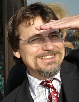 Dr. Bach is Professor, University Eye Hospital, Freiburg, Germany. Visual acuity (VA) is a basic measure of visual function. Bach demonstrated how signal detection theory aids in assessing VA, e.g. in the Freiburg Acuity test. For patients who cannot or will not fully cooperate, visual evoked potentials provide non-invasive objective assessment of VA. We have advanced the methodology embodying signal statistics, Fourier techniques, and fully automatic algorithms to a state where we approach subjective acuity within ± 1 octave in 95% of the cases. Plans were outlined to address the question to what degree subjective perception coincides with the visual resolution of V1. His presentation was “Objective Assessment of Visual Acuity.”