Humeral lengthening using a lower extremity device
An 8-year-old patient’s left proximal humerus unicameral bone cyst treatment took place at Boston Children’s Hospital starting in 2009. As a result of the cyst and subsequent bone grafting, he developed a growth arrest. By age 18, the patient’s left arm was 14 centimeters shorter than his right. In 2019 and 2020, when the patient was 18 and 19 years old, orthopedic surgeon Carley Vuillermin, MBBS, MPH, FRACS, used the PRECICE internal limb-lengthening system to lengthen the patient’s left humerus by five centimeters each summer for a total lengthening of 10 centimeters.
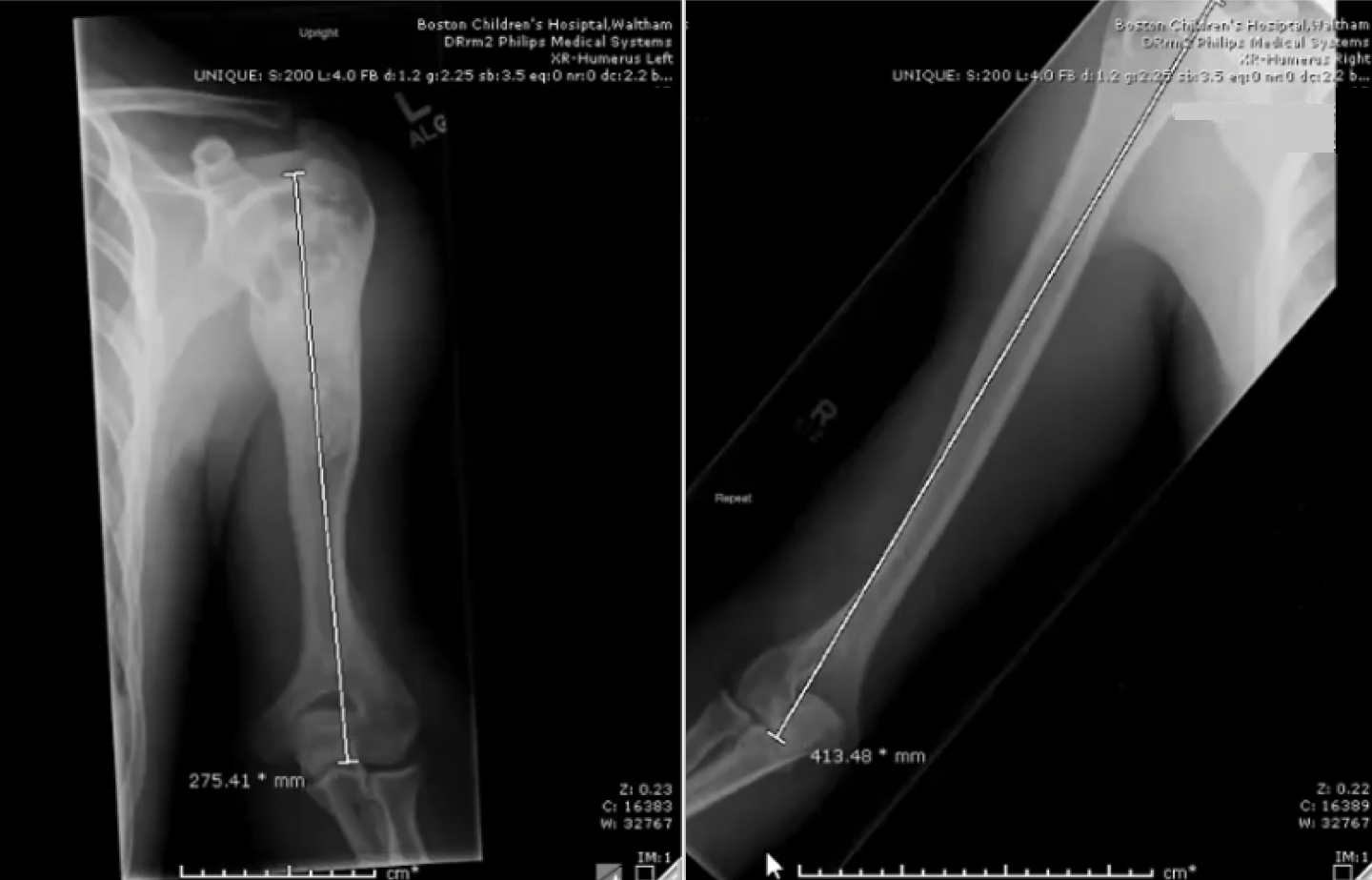
In February 2019 the 18-year-old patient had a 138-millimeter length difference between his left and right arms. The upper left humerus shows the traces of a bone graft performed in 2009 to remove a bone cyst.
Limited options for limb lengthening in the upper extremities
Recent years have brought promising advances in technologies to correct limb-length discrepancies in the lower extremities. The PRECICE nail, an implanted intramedullary device with an externally triggered magnetic lengthening mechanism, offers a number of demonstrated benefits over prior devices, including reliable lengthening, lower rates of infection, and more rapid return to physical activity compared to external devices and earlier mechanical internal lengthening devices.
No internal lengthening device is commercially available specifically for the upper extremity. Reasons for this include the smaller diameter of the bones and lower number of arm-lengthening procedures performed each year. The PRECICE nail is the first device small enough to fit inside the humerus of a skeletally mature patient. The successful use of the tibial and femoral PRECICE system in humeral lengthening in this and a handful of other cases has expanded options for upper extremity lengthening now and in the future.
The procedure and considerations for the upper extremities
Vuillermin inserted the PRECICE nail through the proximal humerus and performed a low-energy corticicotomy. The distraction took place gradually, at a rate of one millimeter per day in three divided increments, eight hours apart. Each lengthening session lasted two minutes. During the first lengthening, the patient achieved a lengthening of 5 centimeters, the maximum length supported by the device. He returned to the OR 12 months later, where his PRECICE nail was replaced, and he underwent a second lengthening. This timing allowed lengthening and consolidation over summer vacation, a period of low sports activity for this particular patient.
Because this patient was an otherwise typically developing adolescent who secondarily developed a growth arrest, the surrounding soft tissues tolerated the lengthening very well. The procedure increased the length of the patient’s upper arm by 37 percent. The humerus is known to lengthen well. Compared to lower extremities, which can tolerate an increase in length of 25 percent, it is often possible to lengthen the humerus up to 40 percent in a single distraction episode, depending on the cause of the foreshortened limb. More caution is required with congenital differences, though this technique has been successfully used in that group of patients as well.
Use of the system for humeral lengthening requires sub-specialized skills in limb lengthening and upper extremity surgery. In particular, the surgeon must be comfortable operating in close proximity to major neurovascular structures. To prevent future shoulder pain and protect its function, the surgeon must take particular care to preserve the rotator cuff. When necessary for nail trajectory, the surgeon must open the rotator cuff before inserting the nail and repair it once the nail is inserted. Altered anatomy, such as moderate humerus varus, as seen in this patient, can mean the cuff can be avoided without these extra steps.
The radial nerve must be protected during corticotomy due to its close proximity to the humeral shaft and commonly altered anatomy. Open insertion of the AP cross bolts in the distal humerus is required to protect the musculocutaneous and lateral cutaneous nerves of the forearm, median nerve, and brachial artery. Proximal cross-bolting is performed with guidance and percutaneously, however the surgeon must take extreme care to protect the axillary nerve.
If rotator cuff repair is part of the operation, the patient must protect the shoulder after surgery. Physical therapy is critically important to maintain range of motion. Therapy for the elbow always commences immediately, while therapy for the shoulder begins after rotator cuff healing.
Typically, it takes twice as long as the bone was distracted over to consolidate after a lengthening procedure, however, the internal nature of the device allows for greater functioning and comfort than prior devices during this period. In line with the manufacturer’s recommendations, Vuillermin anticipates removing the PRECICE nail from this patient’s humerus 12 to 18 months after the second implantation surgery.
Although this technique offers significant advantages, it is still limb lengthening and requires due diligence and strict adherence to limb-lengthening principles. To be considered for humeral lengthening with the PRECICE system, a patient’s growth plate at the proximal humerus should have closed. Following the procedure, the patient should be prepared to adhere to their rehabilitation and physical therapy regimen consistently.
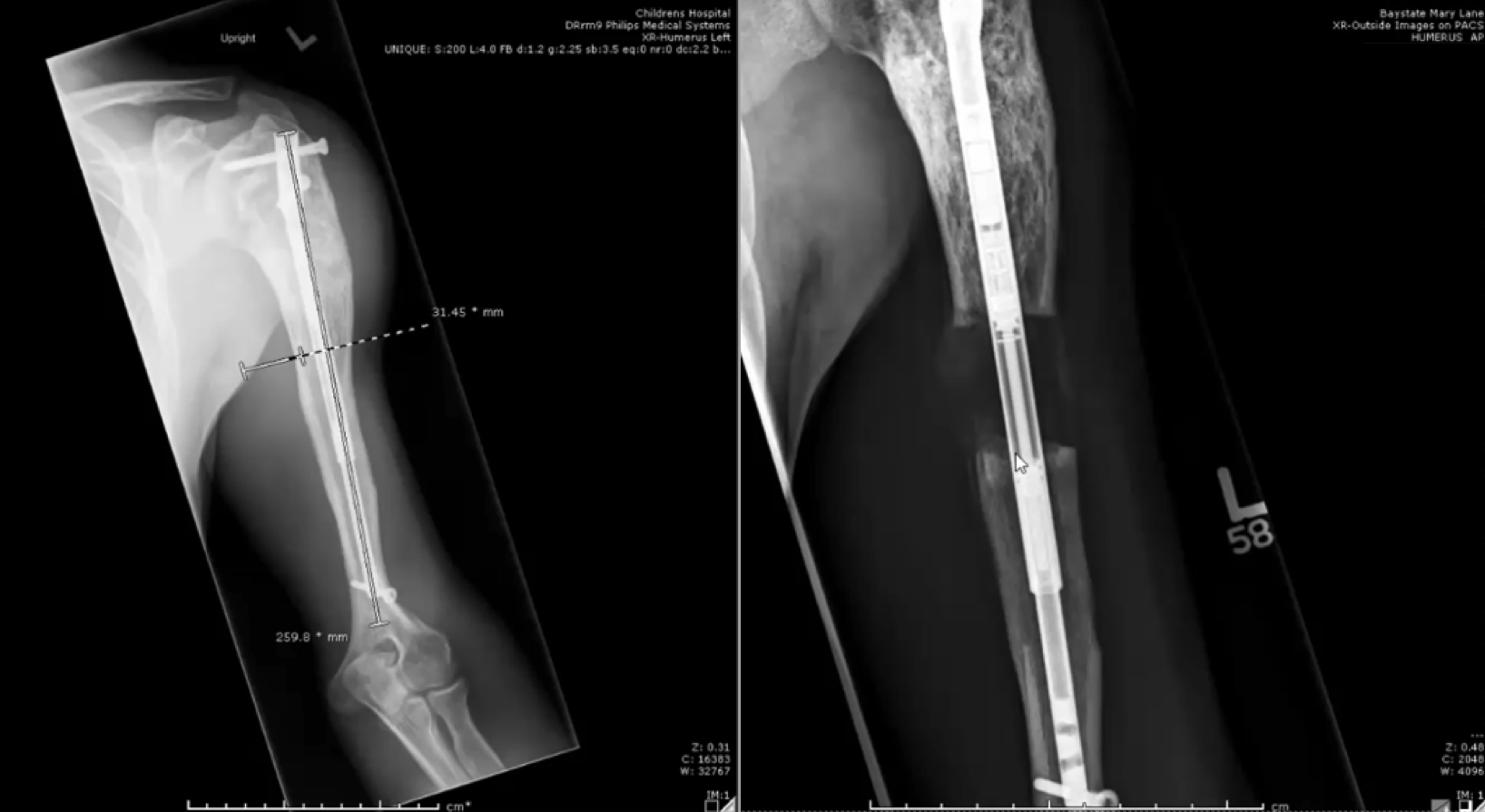
In June 2019, a PRECICE nail was inserted into the left humerus and lengthened by five centimeters; the left image, taken in June 2020, shows where regenerate bone consolidated in the humerus; in July 2020, the bone was lengthened another five centimeters using a second PRECICE nail.
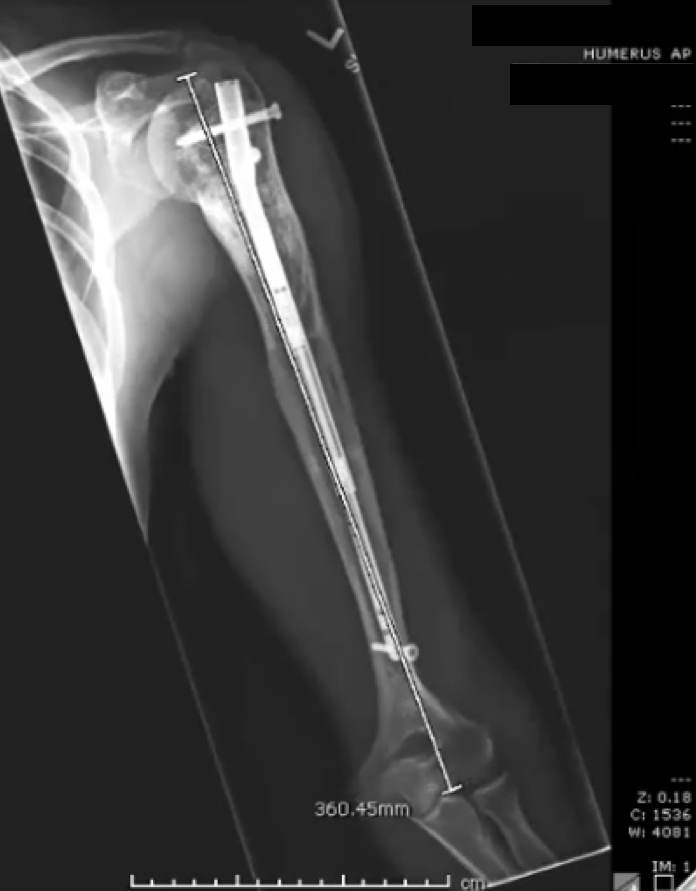
An image taken in January 2021 shows where regenerate bone consolidated in the humerus.